

Multiparametric positron emission tomography/magnetic resonance imaging in nasopharyngeal carcinoma: Correlations between magnetic resonance imaging functional parameters and 18F-fluorodeoxyglucose positron emission tomography imaging biomarkers and their predictive value for treatment failure
- PMID: 33505880
- PMCID: PMC7821831
- DOI: 10.4103/tcmj.tcmj_4_20
Multiparametric positron emission tomography/magnetic resonance imaging in nasopharyngeal carcinoma: Correlations between magnetic resonance imaging functional parameters and 18F-fluorodeoxyglucose positron emission tomography imaging biomarkers and their predictive value for treatment failure
Abstract
Objectives: The clinical significance of positron emission tomography/magnetic resonance imaging (PET/MRI) functional parameters in nasopharyngealcarcinoma (NPC) remains unclear. The purpose of this prospective study was two-fold: (1) to investigate the associations between simultaneously acquired PET/MRI perfusion, diffusion, and glucose metabolism parameters in patients with NPC and (2) to analyze their predictive value with respect to treatment failure.
Materials and methods: We enrolled 85 patients with primary NPC who simultaneously underwent18F-fluorodeoxyglucose PET/CT and PET/MRI before definitive treatment. The following variables were determined: (1) functional parameters from the MRI component, including perfusion values (Ktrans ,kep ,ve , and initial area under the enhancement curve) and apparent diffusion coefficient (ADC) values, and (2) PET parameters, including metabolic tumor volume (MTV). The reciprocal interrelationships between these parameters and their correlations with treatment failure were examined.
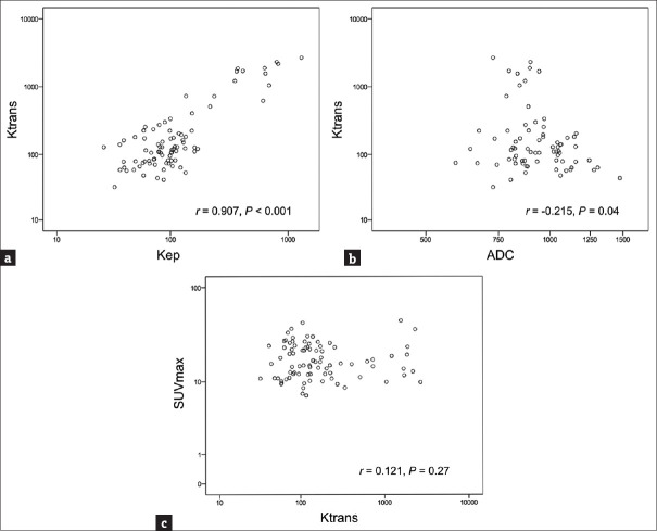
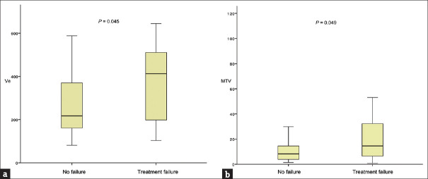
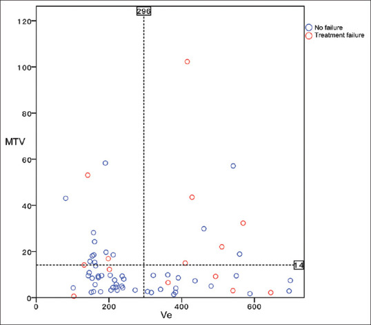
Results: We observed significant negative associations between Ktrans and ADC (r = -0.215, P = 0.049) as well as between ve and ADC (r = -0.22, P = 0.04). Correlations between PET and MRI functional parameters were not statistically significant. Treatment failures were observed in 21.2% of patients without distant metastases. Multivariate analysis identified ve as a significant independent predictor for treatment failure (P = 0.022), whereas MTV showed a borderline significance (P = 0.095). Patients in whom both ve and MTV values were increased had a significantly higher rate of treatment failure (62.5%) than those with either one (21.9%) or no (7.7%) increased parameter (P = 0.004).
Conclusion: Correlation analyses revealed complex interrelationships among PET and MRI indices measured in patients with NPC. These parameters may have a complementary role in predicting treatment failure in this clinical setting.
Keywords: DCE-MRI; Diffusion-weighted MRI; Nasopharyngeal carcinoma; Positron emission tomography/magnetic resonance imaging; Prognosis.
Copyright: © 2020 Tzu Chi Medical Journal.
Conflict of interest statement
There are no conflicts of interest.
Figures
Similar articles
-
Combing MRI Perfusion and 18F-FDG PET/CT Metabolic Biomarkers Helps Predict Survival in Advanced Nasopharyngeal Carcinoma: A Prospective Multimodal Imaging Study.Cancers (Basel). 2021 Mar 28;13(7):1550. doi: 10.3390/cancers13071550. Cancers (Basel). 2021. PMID: 33800542 Free PMC article.
-
Multiparametric functional MRI and 18F-FDG-PET for survival prediction in patients with head and neck squamous cell carcinoma treated with (chemo)radiation.Eur Radiol. 2021 Feb;31(2):616-628. doi: 10.1007/s00330-020-07163-3. Epub 2020 Aug 26. Eur Radiol. 2021. PMID: 32851444 Free PMC article.
-
Dynamic contrast-enhanced magnetic resonance imaging and diffusion-weighted imaging in the activity staging of terminal ileum Crohn's disease.World J Gastroenterol. 2020 Oct 21;26(39):6057-6073. doi: 10.3748/wjg.v26.i39.6057. World J Gastroenterol. 2020. PMID: 33132655 Free PMC article.
-
Gastric cancer and image-derived quantitative parameters: Part 2-a critical review of DCE-MRI and 18F-FDG PET/CT findings.Eur Radiol. 2020 Jan;30(1):247-260. doi: 10.1007/s00330-019-06370-x. Epub 2019 Aug 7. Eur Radiol. 2020. PMID: 31392480 Free PMC article. Review.
-
Prognostic value of pretreatment 18F-FDG PET-CT for nasopharyngeal carcinoma patients.Medicine (Baltimore). 2017 Apr;96(17):e6721. doi: 10.1097/MD.0000000000006721. Medicine (Baltimore). 2017. PMID: 28445287 Free PMC article. Review.
Cited by
-
Comparison of the pre-treatment functional MRI metrics' efficacy in predicting Locoregionally advanced nasopharyngeal carcinoma response to induction chemotherapy.Cancer Imaging. 2021 Nov 10;21(1):59. doi: 10.1186/s40644-021-00428-0. Cancer Imaging. 2021. PMID: 34758876 Free PMC article.
-
Combing MRI Perfusion and 18F-FDG PET/CT Metabolic Biomarkers Helps Predict Survival in Advanced Nasopharyngeal Carcinoma: A Prospective Multimodal Imaging Study.Cancers (Basel). 2021 Mar 28;13(7):1550. doi: 10.3390/cancers13071550. Cancers (Basel). 2021. PMID: 33800542 Free PMC article.
-
Prospective Investigation of 18FDG-PET/MRI with Intravoxel Incoherent Motion Diffusion-Weighted Imaging to Assess Survival in Patients with Oropharyngeal or Hypopharyngeal Carcinoma.Cancers (Basel). 2022 Dec 12;14(24):6104. doi: 10.3390/cancers14246104. Cancers (Basel). 2022. PMID: 36551590 Free PMC article.
References
-
- Chua ML, Wee JT, Hui EP, Chan AT. Nasopharyngeal carcinoma. Lancet. 2016;387:1012–24. - PubMed
-
- O'Sullivan B. Nasopharynx cancer: Therapeutic value of chemoradiotherapy. Int J Radiat Oncol Biol Phys. 2007;69:S118–21. - PubMed
-
- Ng SH, Chan SC, Yen TC, Chang JT, Liao CT, Ko SF, et al. Staging of untreated nasopharyngeal carcinoma with PET/CT: Comparison with conventional imaging work-up. Eur J Nucl Med Mol Imaging. 2009;36:12–22. - PubMed
-
- Zheng D, Yue Q, Ren W, Liu M, Zhang X, Lin H, et al. Early responses assessment of neoadjuvant chemotherapy in nasopharyngeal carcinoma by serial dynamic contrast-enhanced MR imaging. Magn Reson Imaging. 2017;35:125–31. - PubMed
LinkOut - more resources
Full Text Sources
Other Literature Sources