Review
. 2021 Jan 11:10:604124.
doi: 10.3389/fonc.2020.604124. eCollection 2020.

Cancer Stemness: p53 at the Wheel

Affiliations
Review

Cancer Stemness: p53 at the Wheel

Dishari Ghatak et al. Front Oncol. .

Abstract

The tumor suppressor p53 maintains an equilibrium between self-renewal and differentiation to sustain a limited repertoire of stem cells for proper development and maintenance of tissue homeostasis. Inactivation of p53 disrupts this balance and promotes pluripotency and somatic cell reprogramming. A few reports in recent years have indicated that prevalent TP53 oncogenic gain-of-function (GOF) mutations further boosts the stemness properties of cancer cells. In this review, we discuss the role of wild type p53 in regulating pluripotency of normal stem cells and various mechanisms that control the balance between self-renewal and differentiation in embryonic and adult stem cells. We also highlight how inactivating and GOF mutations in p53 stimulate stemness in cancer cells. Further, we have explored the various mechanisms of mutant p53-driven cancer stemness, particularly emphasizing on the non-coding RNA mediated epigenetic regulation. We have also analyzed the association of cancer stemness with other crucial gain-of-function properties of mutant p53 such as epithelial to mesenchymal transition phenotypes and chemoresistance to understand how activation of one affects the other. Given the critical role of cancer stem-like cells in tumor maintenance, cancer progression, and therapy resistance of mutant p53 tumors, targeting them might improve therapeutic efficacy in human cancers with TP53 mutations.

Keywords: GOF mutant p53; cancer stemness; chemoresistance; differentiation; epithelial to mesenchymal transition; miRNAs; therapeutic targeting.


Conflict of interest statement

The authors declare that the research was conducted in the absence of any commercial or financial relationships that could be construed as a potential conflict of interest.

Figures
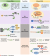
Figure 1
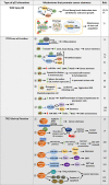
Figure 1
A comparative view of wild-type p53 function in ESC maintenance, differentiation, and somatic-cell reprogramming of human and mouse: ESC maintenance: p53 is maintained in an inactive state in both human and mouse ESCs. In hESCs, deacetylated inactive p53 is present in low levels in the nucleus while in mESCs the inactive p53 protein is abundantly present in the cytoplasm. ESC self-renewal: To ensure ESC self-renewal, p53 is either prevented from entering the nucleus or maintained in an inactive state. In hESCs, Oct4 increases Sirt1 expression which in turn deacetylates p53 and promote its degradation by MDM2. This maintains a low level of p53 in the cell which is crucial to maintain stemness. Endogenous ROS induced p53 nuclear translocation in mESCs is blocked by Sirt1. This prevents p53 mediated suppression of Nanog and stem-cell phenotype is maintained. Phosphorylation and subsequent inactivation of p53 by Aurka also promotes pluripotency of mESCs. ESC differentiation: In hESCs, CBP/p300 mediated acetylation of p53 leads to its activation and subsequent transcription of p21, miR-34a and miR-145 which facilitates differentiation. DNA damage in hESCs also leads to differentiation or apoptosis. When Aurka levels are low in mESCs, p53 transcribes ectodermal and mesodermal genes leading to differentiation. Also, upon DNA damage, p53 primarily promotes differentiation by suppression of Nanog. However, occasionally p53 may also induce anti-differentiation pathway by activating Wnt. Somatic-cell reprogramming: Reprogramming of somatic cells to induced pluripotent stem cells (iPSCs) is primarily inhibited by the p53-p21 pathway in both human and mouse. Additionally, p53 may also induce lincRNAp21 or miR-199a-3p to inhibit reprogramming. The p53-PUMA axis has also been found to suppress reprogramming of mouse embryonic fibroblasts (MEFs).
Figure 2
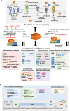
Figure 2
Mechanisms that promote stemness in cancer cells harboring wild-type p53, p53 with loss-of-function mutations or gain-of-function missense mutations: Wild-type p53 modulates the Nanog -Gli positive feedback loop in neural stem cells to control pluripotency. On the contrary, Nanog suppresses p53 activity while Gli activated by Nanog inhibits p53 by activating Mdm2 to promote pluripotency. In hepatic cancer, the stem cell population is maintained by removing mitochondria-associated p53 through mitophagy. TP53 LOF mutations promote various mechanisms that confer stemness phenotype to cancer cells. 1. p53 loss upregulates CD133 which subsequently promotes CSC marker expression and confers stemness. 2. p53 suppresses the cell-surface marker CD44 either by binding to its promoter or by upregulating miR-34a. p53 loss results in increased expression of CD44 and Notch leading to cancer stemness. 3. Loss of p53 also promotes symmetric division of mammary SCs thereby promoting tumorigenesis. 4. Homozygous deletion of p53 in pancreatic acinar cells promotes sphere formation, CSC marker expression as compared to cells with wild type p53. 5. p53 inactivation strongly cooperates with oncogenic Kras mutation in myeloid progenitor cells to induce aggressive AML. 6. p53 loss may also derepress SC marker Nestin to promote differentiation in mature hepatocytes. 7. p53 induces epithelial differentiation by activation of miR-200c. Loss of p53, leads to decreased miR-200c levels and increased expression of its target genes leading to EMT and stemness. TP53 GOF mutations promote cancer stemness by regulating several pathways. 1. Mutant p53 can directly activate CSC markers such as ALDHA1, CD44, and LGR5 to promote stemness. 2. It may regulate Wasp-interacting protein (WIP) that regulates YAP/TAZ stability. 3. Mutant p53 can also promote self-renewal of breast cancer cells by inducing nuclear localization of YAP/TAZ by activating mevalonate pathway. 4. Mutant p53 transcriptionally represses miR-130b and miR- 194, the negative regulators of Zeb1 and Bmi1 respectively, to promote EMT and stemness 5. p53-R273H upregulates lncRNAs, lnc273-31, and lnc273-34 implicated in EMT and CSC maintenance in colorectal cancer cells. 6. GOF mutant p53 promotes typical CSC features of enhanced drug-resistance and prolonged survival by upregulating multidrug resistance gene MDR1, anti-apoptotic genes Bcl-2 and Bcl-xL, and inhibiting pro-apoptotic genes Bax, Bid, and Bad.
Figure 3
Figure 3
(A) Molecular mechanisms of mutant p53 mediated deregulation. The upper panel depicts the upstream signaling pathways deregulated by mutant p53 to promote oncogenesis. The middle panel portrays the different transcription factors, cofactors, and other proteins to which mutant p53 may interact to either enhance or inhibit their binding to the target gene promoter. The lower panel shows the transcriptional and epigenetic targets of mutant p53 classified according to the phenotype they alter. (B) Upstream signals that regulate mutant p53. The upper panel shows the various post translational modifications and chaperons that regulate mutant p53 stability. The modified residues if known, have been mentioned. In others it is not-specified (NS). The lower panel shows the residues in the mutant p53 protein where post-translational modifications occur. Drugs that target interacting proteins of mutant p53, downstream pathways and upstream regulators have been indicated in red in both panels (A, B).

Similar articles

Cited by

References

    1. Chen J. The Cell-Cycle Arrest and Apoptotic Functions of p53 in Tumor Initiation and Progression. Cold Spring Harbor Perspect Med (2016) 6:a026104–a026104. 10.1101/cshperspect.a026104 - DOI - PMC - PubMed
    1. Koifman G, Aloni-Grinstein R, Rotter V. p53 balances between tissue hierarchy and anarchy. J Mol Cell Biol (2019) 11:553–63. 10.1093/jmcb/mjz022 - DOI - PMC - PubMed
    1. Olivier M, Hollstein M, Hainaut P. TP53 mutations in human cancers: origins, consequences, and clinical use. Cold Spring Harbor Perspect Biol (2010) 2:a001008–a001008. 10.1101/cshperspect.a001008 - DOI - PMC - PubMed
    1. Freed-Pastor WA, Prives C. Mutant p53: one name, many proteins. Genes Dev (2012) 26:1268–86. 10.1101/gad.190678.112 - DOI - PMC - PubMed
    1. Sabapathy K, Lane DP. Therapeutic targeting of p53: all mutants are equal, but some mutants are more equal than others. Nat Rev Clin Oncol (2018) 15:13–30. 10.1038/nrclinonc.2017.151 - DOI - PubMed