Review
. 2021 Jan 11:8:620774.
doi: 10.3389/fcell.2020.620774. eCollection 2020.

Closing the Gap: Mechanisms of Epithelial Fusion During Optic Fissure Closure

Affiliations
Review

Closing the Gap: Mechanisms of Epithelial Fusion During Optic Fissure Closure

Brian Ho Ching Chan et al. Front Cell Dev Biol. .

Abstract

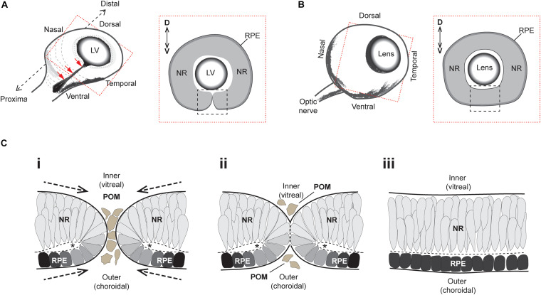
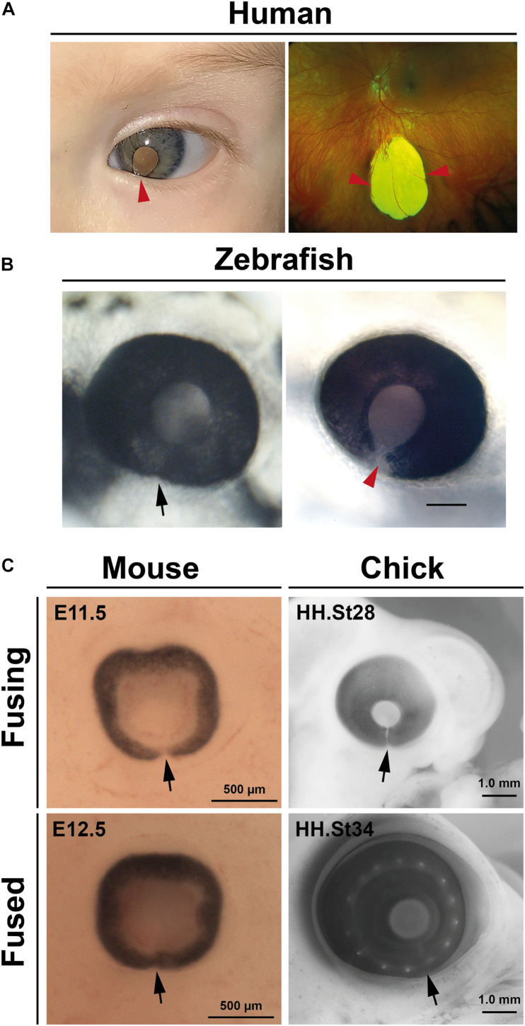
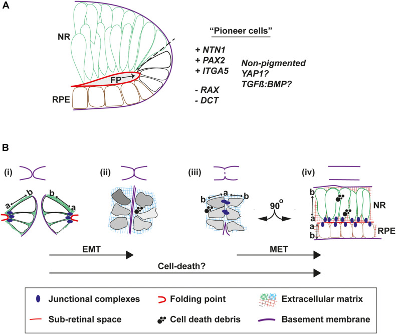
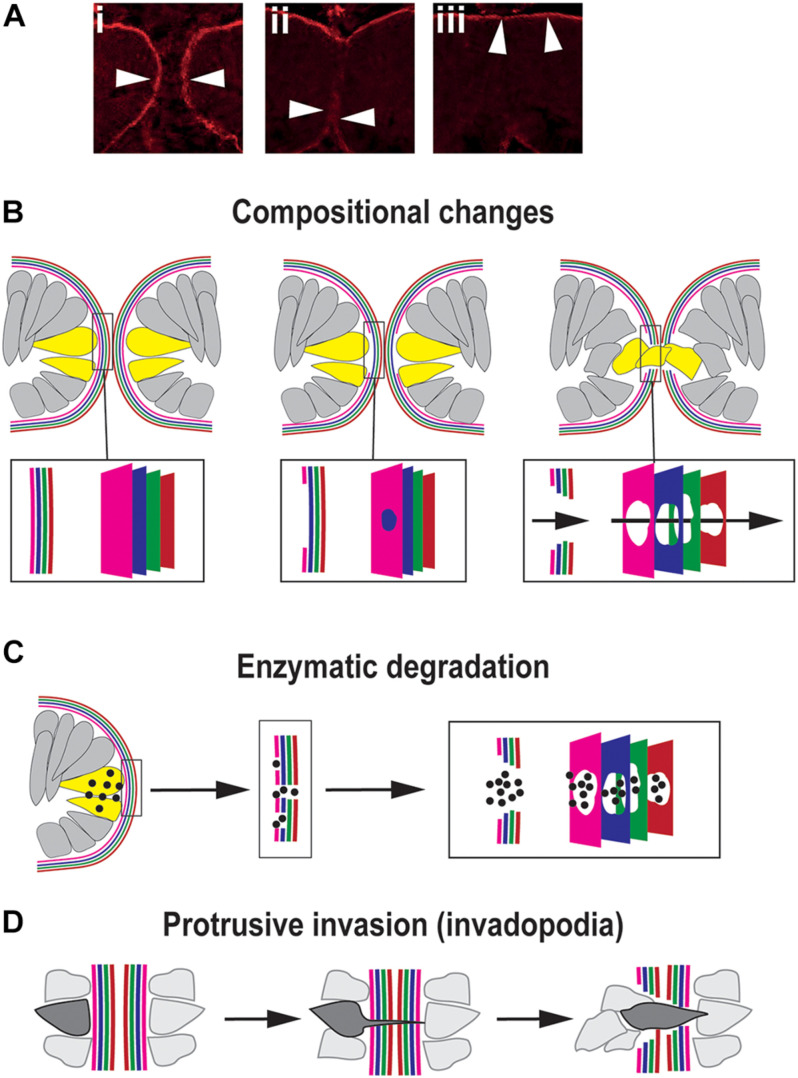
A key embryonic process that occurs early in ocular development is optic fissure closure (OFC). This fusion process closes the ventral optic fissure and completes the circumferential continuity of the 3-dimensional eye. It is defined by the coming together and fusion of opposing neuroepithelia along the entire proximal-distal axis of the ventral optic cup, involving future neural retina, retinal pigment epithelium (RPE), optic nerve, ciliary body, and iris. Once these have occurred, cells within the fused seam differentiate into components of the functioning visual system. Correct development and progression of OFC, and the continued integrity of the fused margin along this axis, are important for the overall structure of the eye. Failure of OFC results in ocular coloboma-a significant cause of childhood visual impairment that can be associated with several complex ocular phenotypes including microphthalmia and anterior segment dysgenesis. Despite a large number of genes identified, the exact pathways that definitively mediate fusion have not yet been found, reflecting both the biological complexity and genetic heterogeneity of the process. This review will highlight how recent developmental studies have become focused specifically on the epithelial fusion aspects of OFC, applying a range of model organisms (spanning fish, avian, and mammalian species) and utilizing emerging high-resolution live-imaging technologies, transgenic fluorescent models, and unbiased transcriptomic analyses of segmentally-dissected fissure tissue. Key aspects of the fusion process are discussed, including basement membrane dynamics, unique cell behaviors, and the identities and fates of the cells that mediate fusion. These will be set in the context of what is now known, and how these point the way to new avenues of research.

Keywords: EMT—epithelial to mesenchymal transition; apoptosis; basement membrane; cell polarity and adhesion; coloboma; eye development; optic fissure closure; transcriptome (RNA-seq).


Conflict of interest statement

The authors declare that the research was conducted in the absence of any commercial or financial relationships that could be construed as a potential conflict of interest.

Figures

FIGURE 1
FIGURE 1
Superficial processes of optic fissure closure. (A) Left: Cartoon of the early 3D optic cup with a ventral cleft—the optic fissure—running along the entire proximal-distal axis (red arrows). Right: Slice-section cut perpendicular to the P-D axis and at the midpoint (hatching in Left) of the optic cup depicting the optic fissure margins (black box) which at this stage are apposed but not fused. (B) Left: Later staged optic cup with a now fully fused optic fissure. Right: Slice-section shows the fused optic fissure (black box) has completed the circumferential continuity of both neural retina and the RPE in the ventral optic cup. (C) Sequence of key cellular and morphological events during optic fissure closure: (i) The nasal and temporal OFMs move toward each other and become spatially apposed, with only a few POM cells separating the edges of each. At this stage, the OFMs are each fully encapsulated by single basement membranes (solid black lines) and the sub-retinal space is folded over as folding points (FP, asterisks); (ii) contact occurs between the two OFMs and POM cells are excluded as the apposed OFMs establish a fusion plate and the separating basement membranes are displaced (thick hatched black lines). (iii) Fusion has occurred and the RPE and NR are each organized into epithelial continuum, while two distinct basement membranes are now established at the inner and outer regions of the optic cup. The FP has become the sub-retinal space (Hatched line). Key- LV, lens vesicle; D-V, dorsal-ventral; NR, neural retina; RPE, retinal pigmented epithelium; POM, periocular mesenchyme.
FIGURE 2
FIGURE 2
Coloboma and optic fissure closure. (A) Human colobomas. Anterior segment photo of the left eye showing iris coloboma and white reflex through the pupil (left) and Ultrawidefield color fundus imaging of the left retina showing an inferior chorioretinal coloboma not involving the optic disc or macula (right). Arrowheads indicate affected regions. (B) Fused optic fissure in zebrafish (left; arrow) and persistent colobomatous defect at 56 hpf (right; arrowhead). (C) Optic fissures (arrows) in the eyes of mouse and chick embryos at fusing and fused stages.
FIGURE 3
FIGURE 3
Cell behaviors that mediate fusion in the OF. (A) Pioneer cells are located at the edges of the fissure margins in a position intermediate between the RPE and neural retina cells. They are negative for the neural retina marker Rx:GFP and for the RPE marker DCT, but are predicted to be positive for NTN1, PAX2, and Integrin-5A. They also remain unpigmented throughout fusion. (B) Morphologies and behaviors of pioneer cells throughout fusion. Top, cartoons to depict progression of fusion. Bottom, (i) Just prior to fusion, pioneer cells at the edge of the fissure margins are tightly anchored to the folding point through junctional complexes at their apical sides, with clear apical-basal (a-b) polarity. (ii) As the margins are tightly apposed their BM come into close contact. Cells lose their a-b polarity and become mesenchymal in appearance, possibly triggered by compositional changes to the ECM or physical forces. (iii) As the BM is removed and cells on either side make contact, transient junctional complexes are present between the apical aspects of pioneer cells. (iv) As fusion is completed, cells reorientate through 90o and align their junctions to the sub-retinal space. Debris from cell death is a feature frequently observed during normal OFC in chick and mouse, but not in zebrafish fissures. Key: EMT, epithelial to mesenchymal transition; MET, mesenchymal to epithelial transition; NR, neural retina; RPE, retinal pigmented epithelia; a, apical; b, basal; FP, folding point.
FIGURE 4
FIGURE 4
Mechanisms for traversing the basement membrane during OFC. (A) Dynamic changes to the localization of the BM (arrowheads) during chick OFC, as defined by antibody staining to laminin (β1). In (i) the OF edges are completely encapsulated by BM, in (ii) the BM has been locally removed or displaced at the fusion plate and cells are mixing between the two margins, and in (iii) the BM is now visible at the inner limits of the neural retina and the outer limit beneath the RPE. Adapted with permission from Hardy et al. (2019). (B) Compositional changes to the BM driven by transcriptional changes in cells at the edges of the OFM. 4x colors are used to depict the four components of BMs: collagen, laminin, HSPG and nidogens, however, the correct orientation or alignment of these in the OF-BM are not known. (C) Enzymatic degradation occurs through local transcription of diffusible proteases (spots) of the MMP or ADAMTS classes expressed by cells (yellow) at the edges of the fissure proximal to BM breaching. (D) Protrusive invasion through intact BM by actin-rich cellular protrusions.

Similar articles

Cited by

References

    1. Abouzeid H., Boisset G., Favez T., Youssef M., Marzouk I., Shakankiry N., et al. (2011). Mutations in the SPARC-related modular calcium-binding protein 1 gene, SMOC1, cause waardenburg anophthalmia syndrome. Am. J. Hum. Genet. 88 92–98. 10.1016/j.ajhg.2010.12.002 - DOI - PMC - PubMed
    1. ALSomiry A. S., Gregory-Evans C. Y., Gregory-Evans K. (2019). An update on the genetics of ocular coloboma. Hum. Genet. 138 865–880. 10.1007/s00439-019-02019-3 - DOI - PubMed
    1. Barbieri A. M., Lupo G., Bulfone A., Andreazzoli M., Mariani M., Fougerousse F., et al. (1999). A homeobox gene, vax2, controls the patterning of the eye dorsoventral axis. Proc. Natl. Acad. Sci. U. S. A. 96 10729–10734. 10.1073/pnas.96.19.10729 - DOI - PMC - PubMed
    1. Bernstein C. S., Anderson M. T., Gohel C., Slater K., Gross J. M., Agarwala S. (2018). The cellular bases of choroid fissure formation and closure. Dev. Biol. 440 137–151. 10.1016/j.ydbio.2018.05.010 - DOI - PMC - PubMed
    1. Božanić D., Tafra R., Saraga-Babić M. (2003). Role of apoptosis and mitosis during human eye development. Eur. J. Cell Biol. 82 421–429. 10.1078/0171-9335-00328 - DOI - PubMed

LinkOut - more resources