. 2021 Jan 11:8:622308.
doi: 10.3389/fcell.2020.622308. eCollection 2020.

Oral and Palatal Dentition of Axolotl Arises From a Common Tooth-Competent Zone Along the Ecto-Endodermal Boundary

Affiliations

Oral and Palatal Dentition of Axolotl Arises From a Common Tooth-Competent Zone Along the Ecto-Endodermal Boundary

Vladimír Soukup et al. Front Cell Dev Biol. .

Abstract

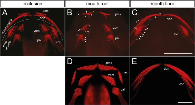
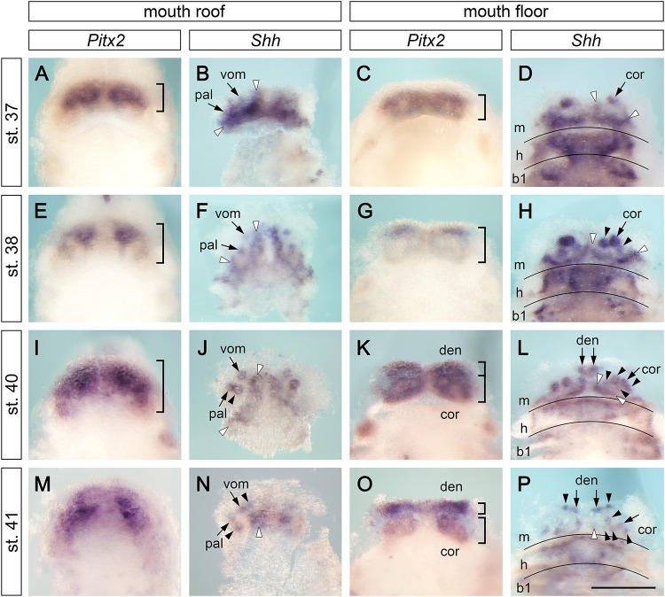
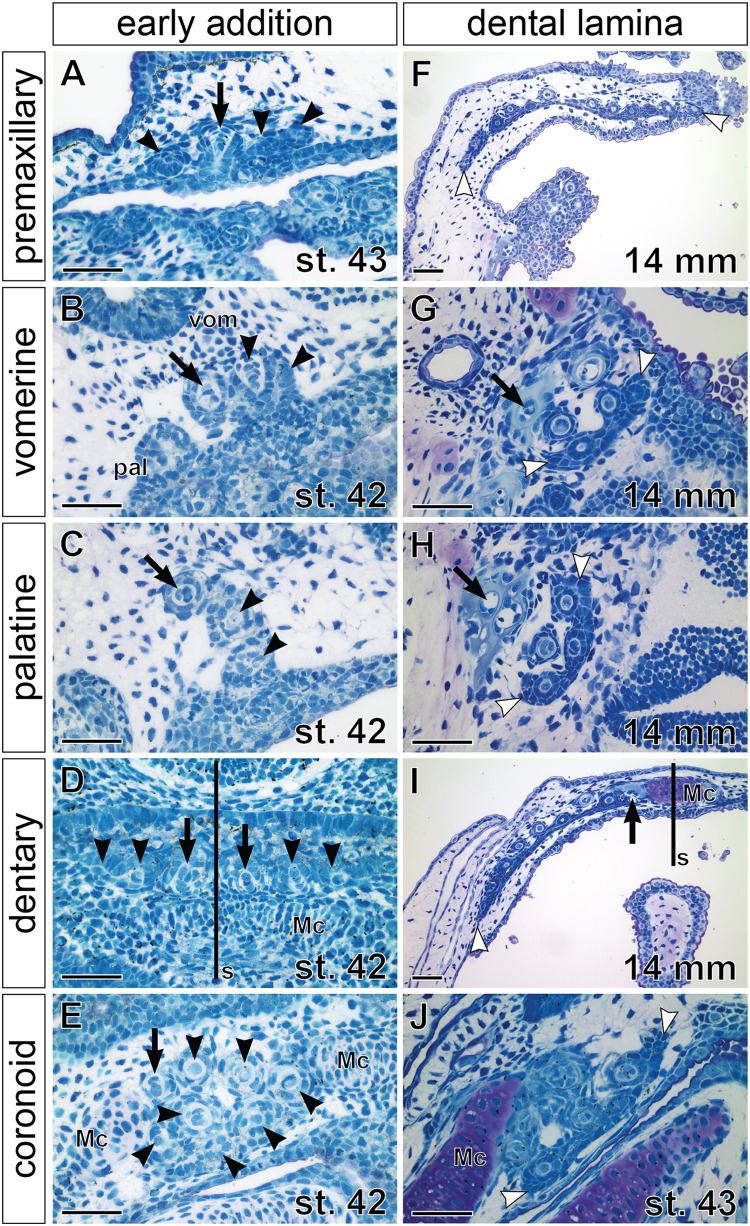
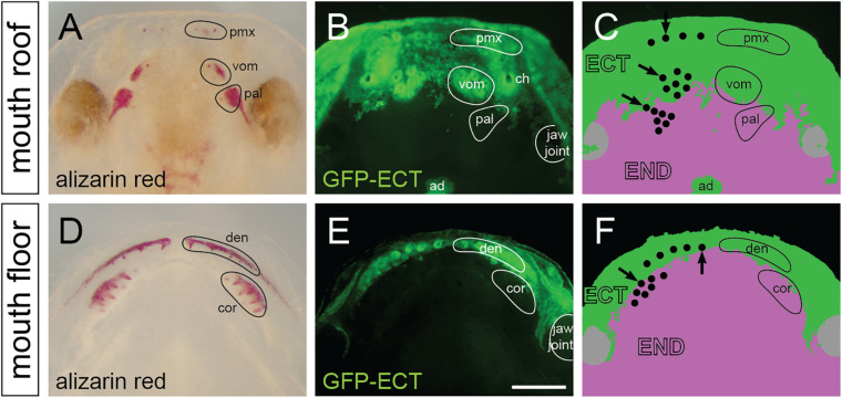
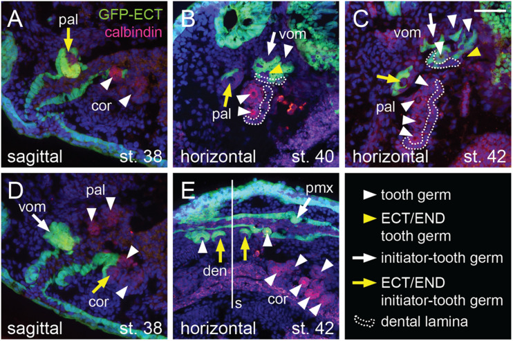
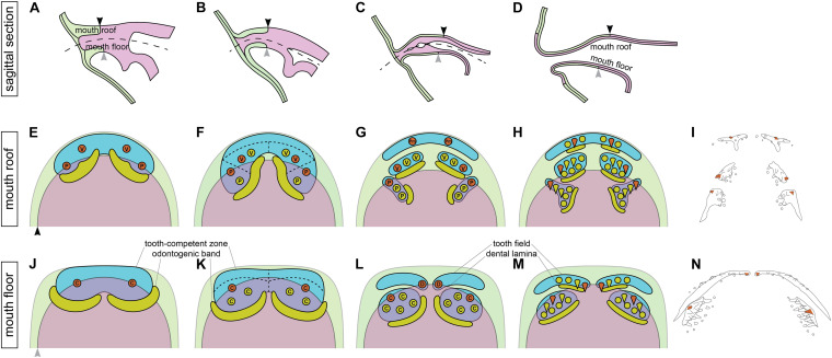
Vertebrate dentitions arise at various places within the oropharyngeal cavity including the jaws, the palate, or the pharynx. These dentitions develop in a highly organized way, where new tooth germs are progressively added adjacent to the initiator center, the first tooth. At the same time, the places where dentitions develop house the contact zones between the outer ectoderm and the inner endoderm, and this colocalization has instigated various suggestions on the roles of germ layers for tooth initiation and development. Here, we study development of the axolotl dentition, which is a complex of five pairs of tooth fields arranged into the typically tetrapod outer and inner dental arcades. By tracking the expression patterns of odontogenic genes, we reason that teeth of both dental arcades originate from common tooth-competent zones, one present on the mouth roof and one on the mouth floor. Progressive compartmentalization of these zones and a simultaneous addition of new tooth germs distinct for each prospective tooth field subsequently control the final shape and composition of the axolotl dentition. Interestingly, by following the fate of the GFP-labeled oral ectoderm, we further show that, in three out of five tooth field pairs, the first tooth develops right at the ecto-endodermal boundary. Our results thus indicate that a single tooth-competent zone gives rise to both dental arcades of a complex tetrapod dentition. Further, we propose that the ecto-endodermal boundary running through this zone should be accounted for as a potential source of instruction factors instigating the onset of the odontogenic program.

Keywords: axolotl; dental arcades; ectoderm; endoderm; initiation; patterning; tooth development.


Conflict of interest statement

The authors declare that the research was conducted in the absence of any commercial or financial relationships that could be construed as a potential conflict of interest.

Figures

FIGURE 1
FIGURE 1
Distribution and composition of axolotl tooth fields. (A) The dentition of an axolotl larva (19 mm TL) is composed of several tooth fields, which are assembled into outer (premaxillary and dentary) and inner dental arcades (vomerine, palatine, and coronoid). These arcades show various field-specific arrangements. Outer arcade fields form initially a single tooth row; inner arcade fields constitute tooth patches. (B,C) Separation of mouth roof from floor shows presence of the oldest tooth (arrow) and addition of new tooth germs (arrowheads) within each field. (B) In the premaxillary field (pmx), new germs are added laterally and medially as the field stretches along the upper jaw, and new germs are also added posteriorly. Vomerine (vom) and palatine (pal) fields develop by addition of new germs medially from the oldest teeth located laterally. (C) In the dentary field (den), the oldest tooth is present close to the mandibular symphysis and new germs are added laterally along the lower jaw, thus forming the tooth row. New germs are further initiated lingually. In the coronoid (cor) field, the oldest tooth is located labially and new germs are added lingually in a hand fan-shaped manner. (D,E) The dentition of a 41 mm TL axolotl larva shows a single-rowed arrangement of premaxillary (pmx), maxillary (max), and dentary (den) fields, and a multi-rowed arrangement of vomerine (vom), palatine (pal), and coronoid (cor) fields. Scale bars equal 1 mm.
FIGURE 2
FIGURE 2
Expression patterns of Pitx2 and Shh at early stages of axolotl odontogenesis. Expression patterns of early odontogenic markers Pitx2 and Shh on the mouth roof and floor whole mounts show initial stages of development of axolotl dentition (in case of Shh expression, the epithelium of the mouth roof was dissected from whole mounts to circumvent masking of the tooth-restricted epithelial signal by the strong Shh signal from the adjacent neural tube). At stages 37–41, Pitx2 labels tooth-competent regions while Shh is restricted to individual tooth germs and into postero-medially placed odontogenic bands (visualized as strands of Shh expression demarcated by white arrowheads). Positions of tooth germs are visible as regions of lower Pitx2 expression. (A,E,I,M) Expression pattern of Pitx2 on the mouth roof is initially restricted to its anterior part but becomes broader at later stages (parentheses). (B,F,J,K) Focal expression of Shh illustrates sequential addition of tooth germs starting from the initiator-teeth of vomerine (vom) and palatine (pal) teeth (B,F, arrows). New tooth germs are added postero-medially in the palatine tooth field (J, black arrowheads) and antero-medially in the vomerine tooth field (N, black arrowheads). (C,G,K,O) Expression pattern of Pitx2 on the mouth floor enlarges posteriorly throughout development and, at stages 40–41, the pattern separates into the prospective dentary (den) and coronoid (cor) tooth fields (K,O, parentheses). (D,H,L,P) Focal Shh expression on the mouth floor marks positions of initiator-teeth of the coronoid (D,H, arrows) and dentary fields (L,P, arrows) and the addition of new tooth germs within both tooth fields (H,L,P, black arrowheads). New tooth germs are added lingually to the initiator tooth germ in the coronoid tooth field (H,L,P, black arrowheads) and laterally from the medially positioned initiator tooth in the dentary tooth field (P, arrowheads). b1, first branchial arch; cor, coronoid tooth field; den, dentary tooth field; h, hyoid arch; m, mandibular arch; pal, palatine tooth field; vom, vomerine tooth field. Scale bar equals 500 μm.
FIGURE 3
FIGURE 3
Expression pattern of Pitx2 at later stages of axolotl odontogenesis. Pitx2 expression becomes gradually restricted to individual tooth germs (arrows mark initiator-tooth germs of respective tooth fields and arrowheads mark sequentially added tooth germs). (A,C,E) Within the mouth roof epithelium, the initial Pitx2 expression delineating individual tooth fields (black broken lines in A) becomes focused into individual tooth germs, inter-germ regions, and the forming successional dental laminae of vomerine (vom) and palatine (pal) tooth fields (C,E). Pitx2 expression is further associated with the developing premaxillary tooth field (pmx), although individual tooth germs cannot be discerned. (B,D,F) Within the mouth floor epithelium, strong Pitx2 expression is visible in the developing tooth germs of dentary (den) and coronoid (cor) tooth fields. cor, coronoid tooth field; den, dentary tooth field; pal, palatine tooth field; pmx, premaxillary tooth field; vom, vomerine tooth field. Scale bars equal 500 μm (scale bar in B is valid for A,B, scale bar in F is valid for C–F).
FIGURE 4
FIGURE 4
Histological analysis of tooth addition within axolotl tooth fields. Horizontal sections (left sides shown, anterior to the top) through individual tooth fields show positions of the respective initiator-tooth (black arrows) and the newly added successive tooth germs (black arrowheads) at stages of early addition (A–E) and later morphogenesis (F–J), when successional dental laminae are clearly discernible (demarcated by white arrowheads). (A) Premaxillary tooth field at stage of opened mouth (stage 43) contains three tooth germs (arrowheads) that are added laterally and medially to the initiator-tooth (arrow). (B,C) Vomerine (vom) and palatine (pal) tooth fields at stage 42 are composed of laterally positioned initiator-teeth (arrow) and two adjacent medially positioned tooth germs (arrowheads). (D) Dentary tooth fields are each initiated from the initiator-tooth (arrows) developing close to the mandibular symphysis (s) and new tooth germs (arrowheads) are added laterally along the Meckel’s cartilage (Mc). (E) Coronoid tooth field is composed of a patch of tooth germs (arrowheads) with the initiator-tooth present antero-laterally (arrow). (F) Premaxillary tooth field of a 14 mm TL larva shows arrangement into a tooth row. New tooth germs are added lingually from the successional dental lamina and, as the upper jaw grows, the premaxillary field expands by further addition of tooth germs at lateral and medial edges of the dental lamina (white arrowheads). (G,H) Vomerine and palatine tooth fields containing several teeth arranged into a tooth patch with new germs arising from the lingually positioned successional dental laminae (white arrowheads). Slight difference in composition exists between these tooth fields that can be ascribed due to the addition of first teeth antero-medially to the initiator-tooth (black arrow) in the vomerine field (B) and postero-medially in the palatine field (C), and to the subsequent development of new tooth germs from the lingually placed successional dental laminae (white arrowheads). (I) Dentary tooth field is arranged into the tooth row. New tooth germs are added from the lingually positioned successional dental lamina and laterally (white arrowhead) due to expansion of the growing Meckel’s cartilage (Mc). (J) Coronoid tooth field at a slightly older stage than that of (E) develops from the lingual successional dental lamina (white arrowheads). Scale bars equal 100 μm.
FIGURE 5
FIGURE 5
Distribution and epithelial derivation of teeth in the axolotl oral cavity. (A,D) Whole mount larvae at stage 44 dissected at the jaw joint and stained by alizarin red demonstrate developing teeth assembled into premaxillary (pmx), vomerine (vom), and palatine (pal) tooth fields on the mouth roof (A), and into dentary (den) and coronoid (cor) tooth fields on the mouth floor (D). (B,C,E,F) Epithelial origin of teeth and schematic interpretation of germ-layer distribution in the axolotl oral cavity (note that only the basal epithelial layer is depicted; the apical layer of the oral epithelium is derived from endoderm). Black dots in C and F represent individual teeth, and positions of initiator-teeth of each tooth field are labeled with black arrows. Teeth developing at the ecto-endoderm boundary are found in vomerine, palatine, dentary, and coronoid tooth fields. ad, adenohypophysis; ch, choana (inner nostril); den, dentary teeth; ECT, ectoderm; END, endoderm; pal, palatine teeth; pmx, premaxillary teeth; cor, coronoid teeth; vom, vomerine teeth. Scale bar equals 500 μm.
FIGURE 6
FIGURE 6
Histological analysis of the epithelial origin of the axolotl teeth. Histological analysis shows details of epithelial origin and modes of addition of teeth (marked by anti-calbindin antibody) within the tooth fields. Arrows mark initiator-tooth germs in each tooth field, arrowheads mark sequentially added tooth germs, yellow arrow/arrowhead denotes tooth developing at the ecto-endoderm boundary, and white arrow/arrowhead denotes tooth developing solely from either ectodermal or endodermal epithelium. (A) Sagittal section showing lateral-most palatine tooth developing at the ecto-endoderm boundary (yellow arrow) close to endoderm-derived coronoid tooth germs (white arrowheads). (B) Horizontal section through vomerine and palatine tooth fields. Two vomerine teeth develop in the ectodermal epithelium (white arrow and arrowhead) and one tooth germ is of ecto-endodermal origin (yellow arrowhead). Palatine tooth field is composed of one ecto-endodermal tooth germ labially (yellow arrow) and two endodermal germs (white arrowheads) lingually. Note the just-arising successional dental laminae (broken lines) that are of endodermal origin in case of the palatine field and ecto-endodermal origin in case of the vomerine field. (C) Horizontal section of later stage vomerine and palatine tooth fields showing ecto-endodermal germ in each vomerine and palatine fields (yellow arrow and arrowhead) next to other ectodermal vomerine teeth and endodermal palatine teeth (white arrowheads). Successional dental laminae (broken lines) are of ecto-endodermal origin in vomerine field and endodermal origin in palatine field. (D) Sagittal section medial to that shown in (A) depicting ecto-endodermal coronoid tooth (yellow arrow), which develops at the level of other tooth germs of vomerine, palatine, and coronoid tooth fields (white arrow and arrowheads). (E) Horizontal section showing ecto-endodermal teeth developing medially in the dentary tooth field (yellow arrows) next to other ectodermal teeth of dentary and premaxillary fields and endodermal teeth of the coronoid field (white arrowheads). den, dentary teeth; pal, palatine teeth; pmx, premaxillary teeth; cor, coronoid teeth; s, symphysis; vom, vomerine teeth. Scale bar equals 50 μm.
FIGURE 7
FIGURE 7
Relations between the germ-layer distribution and patterning of the axolotl dentition. During the course of the axolotl mouth development, oral ectoderm and endoderm do not form a sharp boundary, but instead the basal layer of oral ectoderm involutes to constitute the basal layer of the nascent oral epithelium while the solid endoderm forms the apical layer (A–D). As a result, the ecto-endodermal boundary within the basal layer of the mouth roof (black arrowhead) and floor (gray arrowhead) is different from that within the apical layer. The broken line in (A–C) shows the place of the future mouth cavity. Tooth-competent zones become established in the basal epithelial layer of the mouth roof and floor (blue regions in E,F,J,K) and later become separated into the prospective tooth fields (blue regions in G,H,L,M). Tooth germs arise progressively during the process of this separation. Orange circles mark the positions of the initiator-teeth of each prospective tooth field and yellow circles denote positions of successively added tooth germs. Yellow regions mark positions of odontogenic bands at earlier stages (E,F,J,K) and successional dental laminae at later stages (G,H,L,M). Green and magenta regions show the extent of oral ectoderm and endoderm, respectively. Only the basal epithelial layer, i.e., the tooth-forming layer of the double-layered epithelium, is depicted (the black and gray arrowheads in E,J correspond to those in A–D). First, tooth germs of vomerine (V), palatine (P), and coronoid (C) tooth fields appear prior to those of the premaxillary (Pm) and dentary (D) tooth fields. The initiator-teeth of palatine, coronoid, and dentary tooth fields stereotypically arise at the boundary of ectoderm (green) and endoderm (magenta) (orange circles in F,K,L), while the initiator-teeth of the premaxillary and vomerine tooth fields arise from ectoderm (orange circles in E,G). The successive tooth germs arise from ectoderm in the case of premaxillary and dentary tooth fields, from endoderm in the case of palatine and coronoid tooth fields, and from ectoderm or ecto-endoderm in the case of vomerine tooth fields (F–H,K–M). These early events in oro- and odontogenesis lead to the salamander-specific distribution of teeth on the bones of the upper and lower jaws and on the palate (I,N). Orange teeth in (I,N) represent the initiator-teeth of each tooth field. (I,N) are schematized Figures 1B,C, respectively. den, dentary; pal, palatine; pmx, premaxillary; cor, coronoid; vom, vomerine.

Similar articles

Cited by

References

    1. Balic A. M. (2019). Concise review: cellular and molecular mechanisms of regulation of tooth initiation. Stem Cells 37 26–32. 10.1002/stem.2917 - DOI - PubMed
    1. Barlow L. A. (2000). “Taste buds in ectoderm are induced by endoderm: implications for mechanisms governing taste bud development,” in Regulatory Processes in Development, Wenner-Gren International Series, Vol. 76 eds Ollson, Jacobson C. O. (London: Portland Press; ), 185–190.
    1. Barlow L. A., Northcutt R. G. (1995). Embryonic origin of amphibian taste buds. Dev. Biol. 169 273–285. 10.1006/dbio.1995.1143 - DOI - PubMed
    1. Barlow L. A., Northcutt R. G. (1997). Taste buds develop autonomously from endoderm without induction by cephalic neural crest or paraxial mesoderm. Development 124 949–957. - PubMed
    1. Bordzilovskaya N. P., Dettlaff T. A., Duhon S. T., Malacinski G. M. (1989). “Developmental-stage series of axolotl embryos,” in Developmental Biology of the Axolotl, ed. Malacinski G. M. (Oxford: Oxford University Press; ), 201–219.

LinkOut - more resources