. 2021 Jan 11:8:624337.
doi: 10.3389/fcell.2020.624337. eCollection 2020.

Biological Evaluation of Acellular Cartilaginous and Dermal Matrixes as Tissue Engineering Scaffolds for Cartilage Regeneration

Affiliations

Biological Evaluation of Acellular Cartilaginous and Dermal Matrixes as Tissue Engineering Scaffolds for Cartilage Regeneration

Yahui Wang et al. Front Cell Dev Biol. .

Abstract

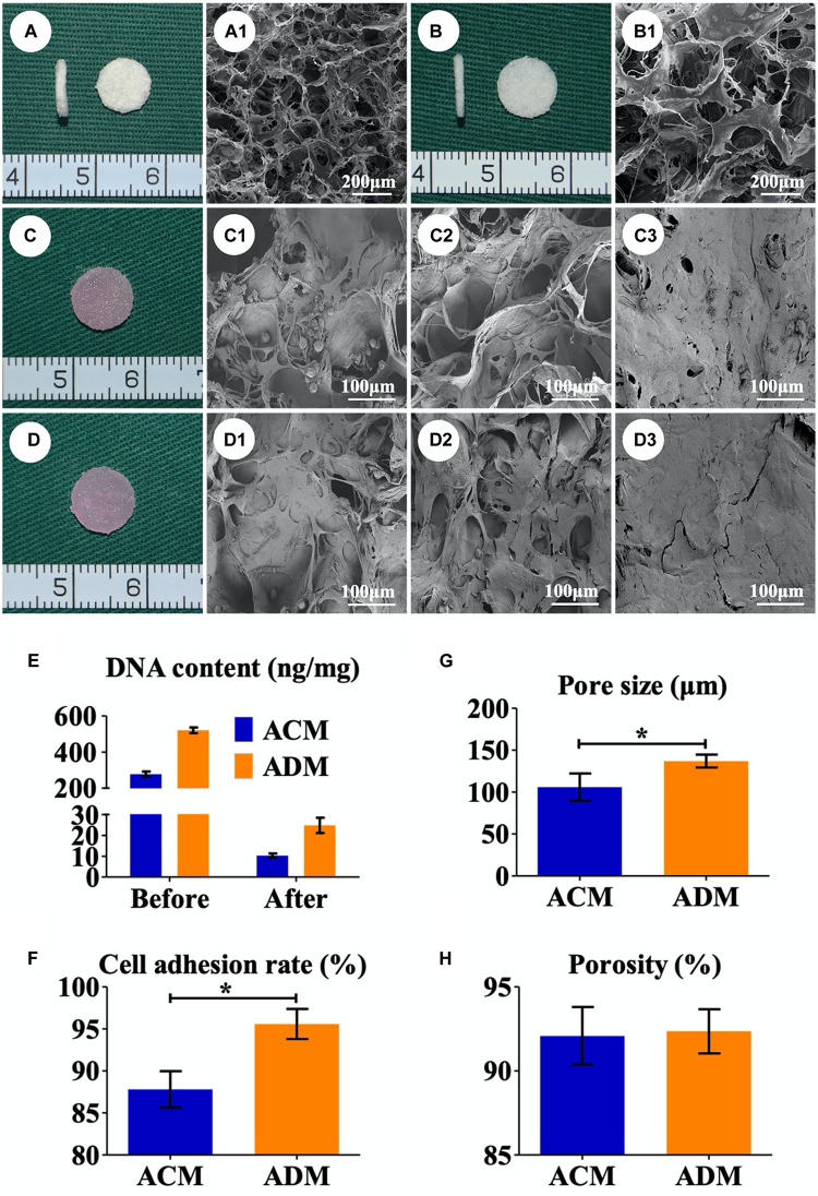
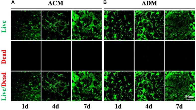
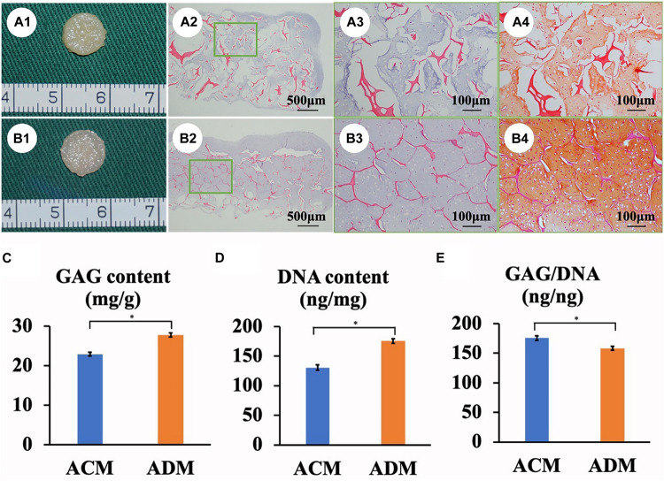
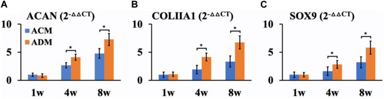
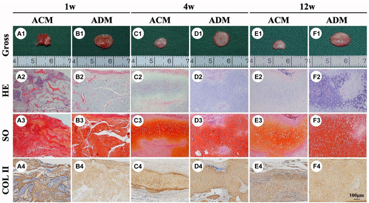
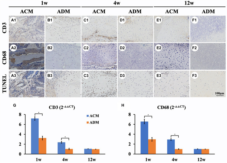
An acellular matrix (AM) as a kind of natural biomaterial is gaining increasing attention in tissue engineering applications. An acellular cartilaginous matrix (ACM) and acellular dermal matrix (ADM) are two kinds of the most widely used AMs in cartilage tissue engineering. However, there is still debate over which of these AMs achieves optimal cartilage regeneration, especially in immunocompetent large animals. In the current study, we fabricated porous ADM and ACM scaffolds by a freeze-drying method and confirmed that ADM had a larger pore size than ACM. By recolonization with goat auricular chondrocytes and in vitro culture, ADM scaffolds exhibited a higher cell adhesion rate, more homogeneous chondrocyte distribution, and neocartilage formation compared with ACM. Additionally, quantitative polymerase chain reaction (qPCR) indicated that expression of cartilage-related genes, including ACAN, COLIIA1, and SOX9, was significantly higher in the ADM group than the ACM group. Furthermore, after subcutaneous implantation in a goat, histological evaluation showed that ADM achieved more stable and matured cartilage compared with ACM, which was confirmed by quantitative data including the wet weight, volume, and contents of DNA, GAG, total collagen, and collagen II. Additionally, immunological assessment suggested that ADM evoked a low immune response compared with ACM as evidenced by qPCR and immunohistochemical analyses of CD3 and CD68, and TUNEL. Collectively, our results indicate that ADM is a more suitable AM for cartilage regeneration, which can be used for cartilage regeneration in immunocompetent large animals.

Keywords: acellular cartilaginous matrix; acellular dermal matrix; cartilage regeneration; immune responses; tissue engineering.


Conflict of interest statement

The authors declare that the research was conducted in the absence of any commercial or financial relationships that could be construed as a potential conflict of interest.

Figures

FIGURE 1
FIGURE 1
Preparation of in vitro ECs. Gross (A) and SEM (A1) images of the ACM scaffold. Gross (B) and SEM (B1) images of the ADM scaffold. Gross views immediately after chondrocytes were seeded onto ACM (C) and ADM (D). SEM images of chondrocyte-ACM constructs after in vitro culture for 1 (C1), 4 (C2), and 7 (C3) days. SEM images of chondrocyte-ADM constructs after in vitro culture for 1 (D1), 4 (D2), and 7 (D3) days. The residual DNA content before and after decellularization in ACM and ADM scaffolds (E). Pore size (G), porosity (H), and cell adhesion rate (F) of ACM and ADM scaffolds. EC, engineered cartilage. *P < 0.05.
FIGURE 2
FIGURE 2
Cell viability of chondrocytes in ACM and ADM scaffolds. Live/dead staining of chondrocytes in ACM for 1, 4, and 7 days (A). Live/dead staining of chondrocytes in ADM for 1, 4, and 7 days (B).
FIGURE 3
FIGURE 3
In vitro ECs formed by chondrocytes seeded in ACM and ADM scaffolds. Gross view, HE staining, and Safranin-O staining of samples in ACM (A1–A4) and ADM (B1–B4) groups after 8 weeks of in vitro culture. Quantitative analysis of the GAG content (C), DNA content (D), and GAG/DNA ratio (E). *P < 0.05.
FIGURE 4
FIGURE 4
QPCR analysis of in vitro ECs in ACM and ADM groups. Expression of ACAN (A), COLIIA1 (B), and SOX9 (C) genes in ACM and ADM groups after 1, 4, and 8 weeks of in vitro culture. *P < 0.05.
FIGURE 5
FIGURE 5
In vivo ECs formed by chondrocytes seeded in ACM and ADM scaffolds. Gross view, HE, Safranin-O, and collagen II immunohistochemical staining of ACM (A1–A4) and ADM (B1–B4) scaffolds after in vivo implantation for 1 week. Gross view, HE, Safranin-O, and collagen II immunohistochemical staining of ACM (C1–C4) and ADM (D1–D4) scaffolds after 4 weeks of in vivo implantation. Gross view, HE, Safranin-O, and collagen II immunohistochemical staining of ACM (E1–E4) and ADM (F1–F4) scaffolds after 12 weeks of in vivo implantation.
FIGURE 6
FIGURE 6
Quantitative analysis of ECs in ACM and ADM groups. Quantitative analysis of the wet weight (A), volume (B), GAG content (C), total collagen (D), collagen II (E), and DNA content (F) in ACM and ADM groups after 1, 4, and 12 weeks of in vivo implantation. *P < 0.05.
FIGURE 7
FIGURE 7
Inflammatory reactions characterized by CD3, CD68, and TUNEL staining. CD3, CD68, and TUNEL staining of in vivo ECs in ACM (A1–A3) and ADM (B1–B3) groups at 1 week after implantation. CD3, CD68, and TUNEL staining of in vivo ECs in ACM (C1–C3) and ADM (D1–D3) groups at 4 weeks after implantation. CD3, CD68, and TUNEL staining of in vivo ECs in ACM (E1–E3) and ADM (F1–F3) groups at 12 weeks after implantation. Expression of CD3 (G) and CD68 (H) genes in ACM and ADM groups after 1, 4, and 12 weeks of in vivo implantation. *P < 0.05.

Similar articles

Cited by

References

    1. Asawa Y., Sakamoto T., Komura M., Watanabe M., Nishizawa S., Takazawa Y., et al. (2012). Early stage foreign body reaction against biodegradable polymer scaffolds affects tissue regeneration during the autologous transplantation of tissue-engineered cartilage in the canine model. Cell Transplant. 21 1431–1442. 10.3727/096368912x640574 - DOI - PubMed
    1. Bayrak A., Pruger P., Stock U. A., Seifert M. (2013). Absence of immune responses with xenogeneic collagen and elastin. Tissue Eng. Part A 19 1592–1600. 10.1089/ten.TEA.2012.0394 - DOI - PMC - PubMed
    1. Ceonzo K., Gaynor A., Shaffer L., Kojima K., Vacanti C. A., Stahl G. L. (2006). Polyglycolic acid-induced inflammation: role of hydrolysis and resulting complement activation. Tissue Eng. 12 301–308. 10.1089/ten.2006.12.301 - DOI - PMC - PubMed
    1. Chen W., Xu Y., Li H., Dai Y., Zhou G., Zhou Z., et al. (2020a). Tanshinone IIA delivery silk fibroin scaffolds significantly enhance articular cartilage defect repairing via promoting cartilage regeneration. ACS Appl. Mater. Interfaces 12 21470–21480. 10.1021/acsami.0c03822 - DOI - PubMed
    1. Chen W., Xu Y., Li Y. Q., Jia L. T., Mo X. M., Jiang G. N., et al. (2020b). 3D printing electrospinning fiber-reinforced decellularized extracellular matrix for cartilage regeneration. Chem. Eng. J. 382:122986 10.1016/j.cej.2019.122986 - DOI