. 2021 Jan 11:7:618660.
doi: 10.3389/fvets.2020.618660. eCollection 2020.

Administration of All-Trans Retinoic Acid to Pregnant Sows Improves the Developmental Defects of Hoxa1-/- Fetal Pigs

Affiliations

Administration of All-Trans Retinoic Acid to Pregnant Sows Improves the Developmental Defects of Hoxa1-/- Fetal Pigs

Haimei Zhou et al. Front Vet Sci. .

Abstract

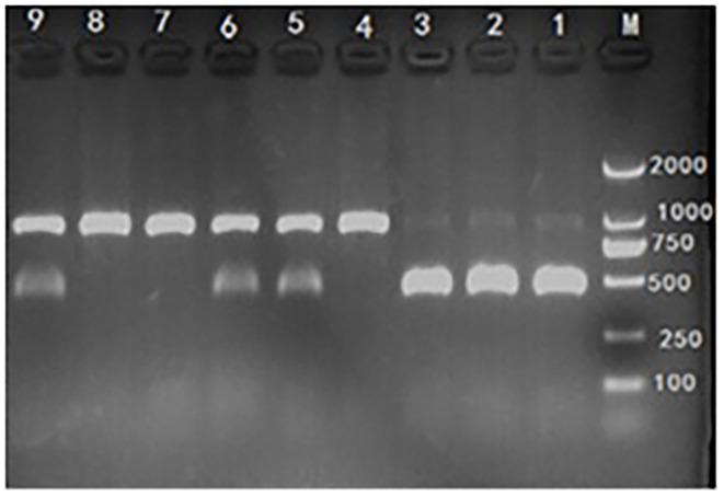
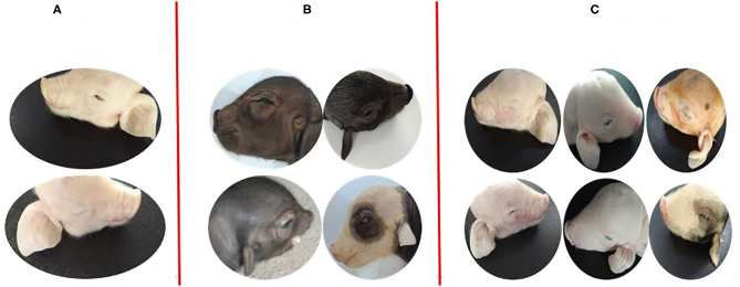
Hoxa1 mutation adversely affect fetal pig development, but whether all-trans retinoic acid (ATRA) administration to Hoxa1+/- pregnant sows can improve Hoxa1-/- fetal pig development defects has not been reported. A total of 24 healthy Hoxa1+/- sows were mated with a healthy Hoxa1+/- boar and randomly assigned to one control group and nine experiment groups. ATRA was orally administered to pregnant sows at the doses of 0, 4, 5, or 6 mg/kg maternal body weight on 12, 13, and 14 days post coitum (dpc), respectively, and a total of 146 live piglets were delivered including 37 Hoxa1-/- piglets and 109 non-Hoxa1-/- piglets. Results indicated that Hoxa1-/- piglets delivered by sows in control group had bilateral microtia, canal atresia and ear's internal defects, and had lower birth liveweight and external ear score than non-Hoxa1-/- neonatal piglets (P < 0.05). Maternal administration with ATRA can effectively correct the development defects of Hoxa1-/- fetal pigs, Hoxa1-/- neonatal piglets delivered by sows administered ATRA at a dose of 4 mg/kg body weight on 14 dpc had higher birth liveweight (P > 0.05) and higher scores of external ear (P < 0.05) compared to Hoxa1-/- neonatal piglets from the control group, but had no significantly difference in terms of birth liveweight and external ear integrity than non-Hoxa1-/- piglets from the control group (P > 0.05). The time of ATRA administration significantly affected Hoxa1-/- fetal development (P < 0.05). Administration of ATRA to Hoxa1+/- pregnant sows at 4 mg/kg body weight on 14 dpc can effectively improve the birth liveweight and ear defects of Hoxa1-/- piglets.

Keywords: Hoxa1 mutation; all-trans retinoic acid; ear defects; fetal pig; intrauterine growth retardation.


Conflict of interest statement

The authors declare that the research was conducted in the absence of any commercial or financial relationships that could be construed as a potential conflict of interest.

Figures

Figure 1
Figure 1
Score criteria for left and right external ear of neonatal piglets. Grade I: normal pinna with ear hole of external auditory canal, score 5. Grade II: small pinna with ear hole of external auditory canal, score 4. Grade III: strip pinna with atresia of external acoustic meatus, score 3. Grade IV: 2-3 “peanut” auricle with atresia of external acoustic meatus, score 2. Grade V: anotia, score 1.
Figure 2
Figure 2
Hoxa1-c.451 G>TC genotyping by polymerase chain reaction-restriction fragment length polymorphism (PCR-RFLP) assay. M: marker. Lane 1, 2, and 3: mutant (Hoxa1−/−), abnormal external ear. Lane 4, 7, and 8: wild type (Hoxa1+/+), normal external ear. Lane 5, 6, and 9: heterozygote (Hoxa1+/−), normal external ear.
Figure 3
Figure 3
Phenotypic comparison of the differences between normal and defected external ears and the differences of repairing defected external ears with ATRA. (A) One non-Hoxa1−/− neonatal piglet from control groups with normal bilateral external ears. (B) The representatives of Hoxa1−/− neonatal piglets from control groups with different defected external ears (C) four Hoxa1−/− neonatal piglets from G7: the left one with the completely repaired bilateral external ears; the middle one and the right two with the partially repaired external ears.
Figure 4
Figure 4
High-resolution CT imaging of internal structures of ears. (A) The non-Hoxa1−/− piglet of Figure 3A had the normal internal structures of ears on each side of the head. (B) The Hoxa1−/− piglet of Figure 3B had the complete absence of EAM, TC and MP. (C) The Hoxa1−/− piglet from the middle of Figure 3C had the partially repaired internal structures of ears. (D) The Hoxa1−/− piglet from the left of Figure 3C had the same normal internal structures of ears as the non-Hoxa1−/− piglet of Figure 3A had. EAM, external auditory meatus. MP, mastoid process. TC, tympanic cavity. AO, auditory ossicle. SCC, semicircular canals. V, vestibule. C, cochlea.

Similar articles

Cited by

References

    1. Bischof JM, Stewart CL, Wevrick R. Inactivation of the mouse Magel2 gene results in growth abnormalities similar to Prader-Willi syndrome. Hum Mol Genet. (2017) 16:2713–9. 10.1093/hmg/ddm225 - DOI - PubMed
    1. Schaaf CP, Gonzalez-Garay ML, Xia F, Potocki L, Gripp KW, Zhang BL, et al. . Truncating mutations of MAGEL2 cause Prader-Willi phenotypes and autism. Nat Genet. (2013) 45:1405–8. 10.1038/ng.2776 - DOI - PMC - PubMed
    1. Guo W, Nie YL, Yan ZQ, Zhu XH, Wang YQ, Guan S, et al. . Genetic testing and pgd for unexplained recurrent fetal malformations with magel2 gene mutation. Sci China Life Sci. (2019) 62:1–9. 10.1007/s11427-019-9541-0 - DOI - PubMed
    1. Cillo C, Cantile M, Faiella A, Boncinelli E. Homeobox genes in normal and malignant cells. J Cell Physiol. (2001) 188:161–9. 10.1002/jcp.1115 - DOI - PubMed
    1. Garcia-Fernández J. The genesis and evolution of homeobox gene clusters. Nat Rev Genet. (2005) 6:881–92. 10.1038/nrg1723 - DOI - PubMed

LinkOut - more resources