

Imaging technologies in the differential diagnosis and follow-up of brown tumor in primary hyperparathyroidism: Case report and review of the literature
- PMID: 33506077
- PMCID: PMC7815655
- DOI: 10.1016/j.bonr.2020.100745
Imaging technologies in the differential diagnosis and follow-up of brown tumor in primary hyperparathyroidism: Case report and review of the literature
Abstract
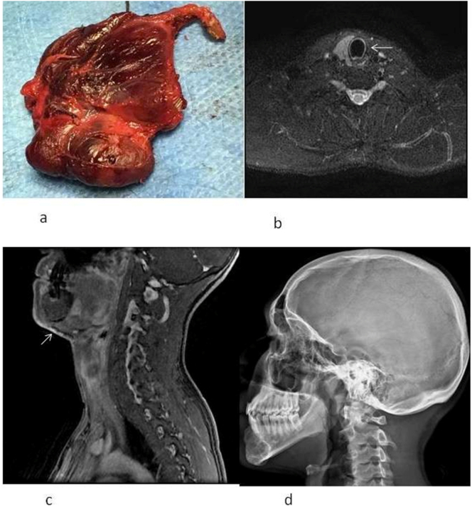
Brown tumors are osteolytic lesions associated with hyperparathyroidism (HPT). They may involve various skeletal segments, but rarely the cranio-facial bones. We report a case of a young boy with a swelling of the jaw secondary to a brown tumor presenting as the first manifestation of primary HPT (PHPT). He was found to have brown tumor located in the skull, as well. Different imaging technologies were employed for the diagnosis and follow-up after parathyroidectomy. We enclose a review of the literature on the employment of such imaging technologies in the differential diagnosis of osteolytic lesions. A multidisciplinary approach comprising clinical, laboratory and imaging findings is essential for the differential diagnosis of brown tumor in PHPT.
Keywords: Brown tumor; MRI; Parathyroid adenoma; Primary hyperparathyroidism.
© 2021 The Authors. Published by Elsevier Inc.
Conflict of interest statement
S.M. served as speaker for Abiogen, Amgen, Bruno Farmaceutici, Diasorin, Eli Lilly, Shire, Sandoz, Takeda. He served in advisory board of Abiogen, Kyowa Kirin, Pfizer, UCB. All other authors declare no conflict of interests.
Figures

References
-
- Argiro R., Diacinti D., Sacconi B., Iannarelli A., Diacinti D., Cipriani C., Pisani D., Romagnoli E., Biffoni M., Di Gioia C., Pepe J., Bezzi M., Letizia C., Minisola S., Catalano C. Diagnostic accuracy of 3T magnetic resonance imaging in the preoperative localisation of parathyroid adenomas: comparison with ultrasound and 99mTc-sestamibi scans. Eur. Radiol. 2018;28:4900–4908. - PubMed
-
- Bilezikian J.P., Bandeira L., Khan A., Cusano N.E. Hyperparathyroidism. Lancet. 2018;391:168–178. - PubMed
-
- Brabyn P., Capote A., Belloti M., Zylberberg I. Hyperparathyroidism diagnosed due to Brown tumors of the jaw: a case report and literature review. Journal of oral and maxillofacial surgery : official journal of the American Association of Oral and Maxillofacial Surgeons. 2017;75:2162–2169. - PubMed
-
- Carpten JD, Robbins CM, Villablanca A, Forsberg L, Presciuttini S, Bailey-Wilson J, Simonds WF, Gillanders EM, Kennedy AM, Chen JD, Agarwal SK, Sood R, Jones MP, Moses TY, Haven C, Petillo D, Leotlela PD, Harding B, Cameron D, Pannett AA, Hoog A, Heath H, 3rd, James-Newton LA, Robinson B, Zarbo RJ, Cavaco BM, Wassif W, Perrier ND, Rosen IB, Kristoffersson U, Turnpenny PD, Farnebo LO, Besser GM, Jackson CE, Morreau H, Trent JM, Thakker RV, Marx SJ, Teh BT, Larsson C, Hobbs MR (2002) HRPT2, encoding parafibromin, is mutated in hyperparathyroidism-jaw tumor syndrome. Nature genetics 32:676–680. - PubMed
-
- Cebesoy O., Karakok M., Arpacioglu O., Baltaci E.T. Brown tumor with atypical localization in a normocalcemic patient. Arch. Orthop. Trauma Surg. 2007;127:577–580. - PubMed
Publication types
LinkOut - more resources
Full Text Sources
Other Literature Sources
