Review
. 2020 Dec 31;24(1):102012.
doi: 10.1016/j.isci.2020.102012. eCollection 2021 Jan 22.

3D-printed microneedles in biomedical applications

Affiliations
Review

3D-printed microneedles in biomedical applications

Sajjad Rahmani Dabbagh et al. iScience. .

Abstract

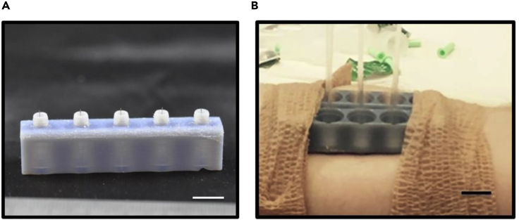
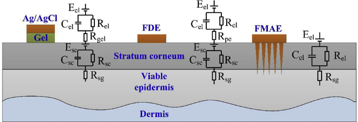
Conventional needle technologies can be advanced with emerging nano- and micro-fabrication methods to fabricate microneedles. Nano-/micro-fabricated microneedles seek to mitigate penetration pain and tissue damage, as well as providing accurately controlled robust channels for administrating bioagents and collecting body fluids. Here, design and 3D printing strategies of microneedles are discussed with emerging applications in biomedical devices and healthcare technologies. 3D printing offers customization, cost-efficiency, a rapid turnaround time between design iterations, and enhanced accessibility. Increasing the printing resolution, the accuracy of the features, and the accessibility of low-cost raw printing materials have empowered 3D printing to be utilized for the fabrication of microneedle platforms. The development of 3D-printed microneedles has enabled the evolution of pain-free controlled release drug delivery systems, devices for extracting fluids from the cutaneous tissue, biosignal acquisition, and point-of-care diagnostic devices in personalized medicine.

Keywords: Biomaterials; Biomedical Materials; Materials in Biotechnology.


Figures

Figure 1
Figure 1
The main purpose of the microneedles (MNs) patch application is to deliver the active biomolecules to the close or remote sites inside the body (A) A cutaneous tissue consists of three layers: epidermis, dermis, and hypodermis. (B) The outermost layer, the epidermis, is a keratinized stratified squamous epithelium with closely packed epithelial cells. The dermis layer lies beneath the epidermis and is composed of connective tissue with vessels and nerves ending. The innermost layer of the skin is so-called hypodermis, which is characterized by the loose connective tissue and the existence of adipocytes.
Figure 2
Figure 2
An illustration of the different types of microneedles (MNs) and delivery means (A) The most common types of MNs are solid, coated, dissolving, and hollow. (B) MNs pass drugs through the outermost layers of skin to deliver desired cargo to the dermis layer, with different approaches (Kim et al., 2012). Adapted with permission from (Kim et al., 2012). Copyright 2013, Elsevier.
Figure 3
Figure 3
Step-by-step illustration of fabricating microneedles (MNs) with stereolithography (SLA) 3D printing and replica mold method (A) The design procedure was followed by 3D printing of the designed structure using an SLA printer. (B) The 3D-printed MNs were washed and then cured with UV light, followed by filling the basin with UV-curable resin to obtain the desired MN height, resulting in a microneedle arrays (MNA) master. In the next step, using silicone, degassing by the vacuum chamber, and heat curing in the oven, the final female master mold can be produced (Krieger et al., 2019). Adapted with permission from (Krieger et al., 2019). Copyright 2019, Springer Nature.
Figure 4
Figure 4
Scanning electron microscope (SEM) images of microneedles (MNs), fabricated by stereolithography (SLA), with different feature sizes (A) Fabricated MNs via various printing layer heights: 100, 50, and 25 micrometers. Although lower layer heights bring about better surface quality, the printing process will be slower. Hence, choosing the proper layer height is a trade-off between surface quality and time. (B) Tip of fabricated MNs (top:100 μm, bottom: 25 μm layer height). (C) Different aspect ratios. A higher aspect ratio here means a gradual increase of shaft thickness that results in a painless insertion of MN. However, thinner needles possess low mechanical strength that can result in breaking the needle in the insertion process. (D) The discrepancy between the input and output height. Higher aspect ratios suffer more from discrepancy issues (Krieger et al., 2019). Adapted with permission from (Krieger et al., 2019). Copyright 2019, Springer Nature.
Figure 5
Figure 5
Scalable microneedle array (MNA) fabrication in six steps, using 3D direct laser printing and molding (A) 3D design of size features of the MNA, based on the proposed application. (B) Direct production of designed MNA using 3D direct laser printing. MNs with a height of 750 μm, base diameter of 150 μm, arrow base radius of 250 μm, and a tip angle of 30° were produced. (C) Replicating the master MNA mold with high fidelity by a two-step micromolding process. (D) Arranging multiple MNA master molds on a 3D printed holder. (E) Production of MNA fabrication molds by polydimethylsiloxane (PDMS). (F) Loading the desired drug on the tip of the fabricated MNAs by a spin-casting method (Balmert et al., 2020). Adapted with permission from (Balmert et al., 2020). Copyright 2020, Elsevier.
Figure 6
Figure 6
Sequential steps of microneedles (MNs) fabrication by magneto-rheological drawing lithography (MRDL), an additive manufacturing method (A) Pillar tips, with a diameter of 700 μm, were coated by dipping in curable magnetorheological fluid (CMRF). (B) Then, the pillars were moved downward, toward a substrate, with a constant speed of 1.5 mm/s to press droplets to the substrate for 1 s. (C) Pillars moved upward with the speed of 1.5 mm/s and stopped in 12 mm from the substrate, resulting in a necking effect. (D and E) When the thinned CMRF lines broke up at room temperature, the MNs were solidified at 100°C for 1 h. (F) Subsequently, the fabricated MNs were coated by titanium (Ti) and gold (Au) using a magnetron sputtering machine (Ren et al., 2017). Adapted with permission from (Ren et al., 2017). Copyright 2017, Elsevier.
Figure 7
Figure 7
The integration of microfluidic devices with microneedles offers better fluid management abilities, resulting in a more advanced level of controlled drug delivery (A) An illustration of the proposed device fabricated by stereolithography 3D printing, with three separate microfluidic inlets and microneedles (MN) as the outlet. The zoomed section shows the MNs. (B) Ex vivo transdermal delivery of three model drugs, from multiple inlets, into a porcine skin (Yeung et al., 2019). Adapted with permission from (Yeung et al., 2019). Copyright 2019, American Institute of Physics.
Figure 8
Figure 8
MNs with a core-shell structure featuring programmable drug-releasing kinetics with a one-time insertion mimicking the multi-injection drug or vaccine delivery systems (A) Schematic view of the three constituent elements of the fabricated MNs: shell, cap, and drug core, demonstrating the underlying principle of the kinetics of controlled drug delivery. (B) Optical image of the fabricated MNs. Scale bar: 0.5 cm. (C) Step-by-step 3D manufacturing process of MNs: (1) a PDMS negative mold of MNs was filled with PLGA; (2) a positive polylactide (PLA) mold was used to encroach the MNs inside the PDMS mold; (3 and 4) arrays of drug cores were aligned and loaded into MNs in a high-throughput drug-loading process; (v) PLA supporting patch including the cap layer was attached to the MNs in a heat-sintering process. (vi) The MNs were ready after peeling off the PDMS mold (Tran et al., 2020). Adapted with permission from (Tran et al., 2020). Copyright 2020, Springer Nature.
Figure 9
Figure 9
Application of the microneedle (MN) patches in cancer therapy (A) The integration of antibodies, adjuvants, peptides, and genetic elements is done to stimulate immune cells and increase tumoricidal outcome along with chemotherapy. (B) Intelligent needle structures are used to control the systemic levels of endocrine hormones upon the onset of metabolic diseases.
Figure 10
Figure 10
Microneedle arrays (MNAs) comprised of 3D-printed holder arrays and ultra-fine needles for dermal interstitial fluid extraction (A) 3D-printed MN holder with attached needles, coated with a silicone lubricant to lessen the insertion pain (scale bar: 1 cm). MN holders not only do affect the extraction rate by applying pressure on the surroundings of the insertion site but they also protect the fragile MN during the insertion process by an unskilled patient. (B) The fabricated MNA being used for dermal interstitial fluid extraction in a human subject. The extracted fluid was collected in the glass capillary tubes (scale bar: 1 cm) (Miller et al., 2018). Adapted with permission from (Miller et al., 2018). Copyright 2018, Springer Nature.
Figure 11
Figure 11
Different layers constituting the skin and circuit model corresponding to different bio-signal recording techniques Whereas utilizing conventional wet electrodes (e.g., Ag/AgCl probes) require the use of gel, causing allergenic reactions in a number of patients, flexible dry electrode (FDE) probes require skin preparation step (e.g., shaving body hairs), prior to signal acquisition, bringing about unwillingness in patients for the test. However, microneedle arrays (MNA) not only do not need any gel usage or skin preparation, but MNs also do not suffer from background noise and high signal-to-noise rate owing to penetrating layers beneath the outermost layer of skin (Ren et al., 2017). Adapted with permission from (Ren et al., 2017). Copyright 2017, Elsevier.

References

    1. Aimar A., Palermo A., Innocenti B. The role of 3D printing in medical applications: a state of the art. J. Healthc. Eng. 2019;2019:5340616. - PMC - PubMed
    1. Aksit A., Arteaga D.N., Arriaga M., Wang X., Watanabe H., Kasza K.E., Lalwani A.K., Kysar J.W. In-vitro perforation of the round window membrane via direct 3-D printed microneedles. Biomed. Microdevices. 2018;20:47. - PMC - PubMed
    1. Ali R., Mehta P., Arshad M., Kucuk I., Chang M., Ahmad Z. Transdermal microneedles—a materials perspective. AAPS PharmSciTech. 2020;21:12. - PubMed
    1. Amin R., Knowlton S., Hart A., Yenilmez B., Ghaderinezhad F., Katebifar S., Messina M., Khademhosseini A., Tasoglu S. 3D-printed microfluidic devices. Biofabrication. 2016;8:022001. - PubMed
    1. Amin R., Knowlton S., Yenilmez B., Hart A., Joshi A., Tasoglu S. Smart-phone attachable, flow-assisted magnetic focusing device. RSC Adv. 2016;6:93922–93931.

LinkOut - more resources