. 2020 Dec 31;24(1):102022.
doi: 10.1016/j.isci.2020.102022. eCollection 2021 Jan 22.

Cryo-EM structure of human mitochondrial HSPD1

Affiliations

Cryo-EM structure of human mitochondrial HSPD1

David P Klebl et al. iScience. .

Abstract

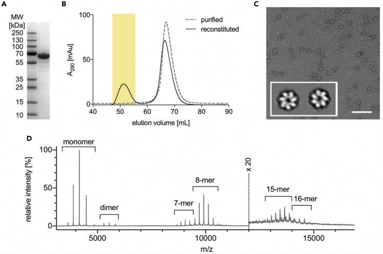
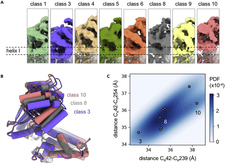
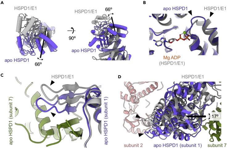
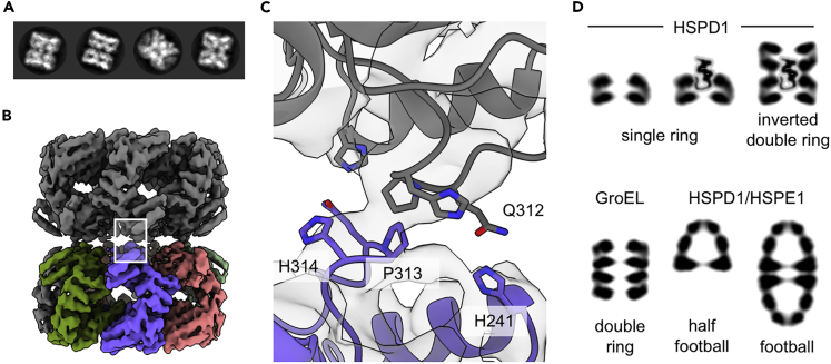
Chaperonins play an important role in folding newly synthesized or translocated proteins in all organisms. The bacterial chaperonin GroEL has served as a model system for the understanding of these proteins. In comparison, its human homolog, known as mitochondrial heat shock protein family member D1 (HSPD1) is poorly understood. Here, we present the structure of HSPD1 in the apo state determined by cryo-electron microscopy (cryo-EM). Unlike GroEL, HSPD1 forms mostly single ring assemblies in the absence of co-chaperonin (HSPE1). Comparison with GroEL shows a rotation and increased flexibility of the apical domain. Together with published structures of the HSPD1/HSPE1 co-chaperonin complex, this work gives insight into the structural changes that occur during the catalytic cycle. This new understanding of HSPD1 structure and its rearrangements upon complex formation may provide new insights for the development of HSPD1-targeting treatments against a diverse range of diseases including glioblastoma.

Keywords: Molecular Biology; Molecular Structure.


Conflict of interest statement

The authors declare no conflicts of interest.

Figures

None
Graphical abstract
Figure 1
Figure 1
Reconstitution and characterization of HSPD1 oligomers (A) SDS PAGE gel for purified HSPD1 monomer. (B) Size exclusion chromatography of purified (gray dashed line) and reconstituted (black solid line) HSPD1 with the peak for oligomeric HSPD1 highlighted in yellow. (C) Negative stain electron micrograph of the oligomeric HSPD1 (scale bar, 100 nm). The inset shows two representative 2D classes. (D) Native mass spectrum of reconstituted HSPD1 showing multiple charge state distributions; different species are labeled. Above 12,000 m/z, the spectral intensity has been multiplied by 20.
Figure 2
Figure 2
Cryo-EM structure of HSPD1 single ring (A) Raw micrograph of apo HSPD1 (scale bar, 50 nm). The bottom panel shows representative 2D classes from the final particle selection. (B) Cryo-EM reconstruction of the apo HSPD1 single ring at 3.5 Å, colored by subunit. (C) Atomic model for apo HSPD1 in the EM density (transparent). On the right, one subunit is shown from the side, and the three domains are labeled.
Figure 3
Figure 3
Conformational variability in the apical domain (A) Eight subunit reconstructions show variable apical domain positions. The position of helix I in class 1 is indicated by dotted lines to help compare the classes. (B) Overlay of flexibly fitted models for classes 3, 8, and 10 in purple, gray, and light red. (C) Plot of measured equatorial/apical domain distances for all eight classes. Each class is represented by a gray point; classes 3, 8 and 10 are labeled. Points approximately lie on the diagonal, showing that the entire apical domain rotates upward. The background color is an estimate of the probability to find a subunit in a certain conformation, taking the particle number in each class into account. PDF stands for probability density function.
Figure 4
Figure 4
Comparing structures of apo HSPD1 and the half-football complex (HSPD17/HSPE17/MgADP) In all panels, the half-football complex is shown in gray (orange and green for neighboring subunits), and the corresponding apo HSPD1 subunit is shown in purple. Arrowheads are used to indicate the regions that show the biggest change. (A) Rotation of the apical domain. Shown are the top (left) and side views of the domain (right). The black line is the rotation axis. (B) Small changes near the nucleotide binding site involving Asp85. (C) Altered position of the inter-subunit beta sheet in apo HSPD1 between subunits 1 and 7. (D) Relative rotation of the equatorial domains in the single ring. The rotation axis is shown as black line.
Figure 5
Figure 5
The HSPD1 inverted double ring (A) Selected 2D classes of the inverted double ring. (B) D7-symmetrical reconstruction of the inverted double ring with one ring colored by subunit and the second ring in gray. The interface between the two rings is highlighted by the white rectangle. (C) Magnified view of the inter-ring interface of the double ring with potential interacting residues labeled. (D) Schematic of the HSPD1 oligomeric states, apo GroEL, and HSPD1/HSPE1 complexes in comparison. Depicted are central sections of side views for the complexes. The eighth subunit is not resolved by cryo-EM, thus probably disordered, and has been placed in the substrate binding site on the apical domains. According to native MS data, the inverted double ring may contain one or two additional (unresolved) subunits.

References

    1. Bartolucci C., Lamba D., Grazulis S., Manakova E., Heumann H. Crystal structure of wild-type chaperonin GroEL. J. Mol. Biol. 2005;354:940–951. - PubMed
    1. Braig K., Otwinowski Z., Hegde R., Boisvert D.C., Joachimiak A., Horwich A.L., Sigler P.B. The crystal structure of the bacterial chaperonin GroEL at 2.8 A. Nature. 1994;371:578–586. - PubMed
    1. Bukau B., Horwich A.L. The Hsp70 and Hsp60 chaperone machines. Cell. 1998;92:351–366. - PubMed
    1. Cappello F., Marino Gammazza A., Palumbo Piccionello A., Campanella C., Pace A., Conway de Macario E., Macario A.J. Hsp60 chaperonopathies and chaperonotherapy: targets and agents. Expert Opin. Ther. Targets. 2014;18:185–208. - PubMed
    1. Chen L., Sigler P.B. The crystal structure of a GroEL/peptide complex: plasticity as a basis for substrate diversity. Cell. 1999;99:757–768. - PubMed