Review
. 2021 Jan 28;12(1):9.
doi: 10.1186/s13244-020-00944-w.

A pictorial review of the utility of CEUS in thoracic biopsies

Affiliations
Review

A pictorial review of the utility of CEUS in thoracic biopsies

Gibran T Yusuf et al. Insights Imaging. .

Abstract

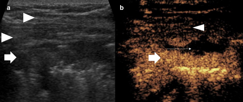
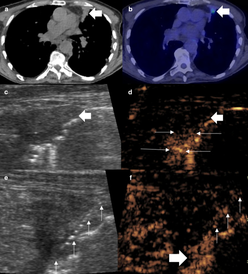
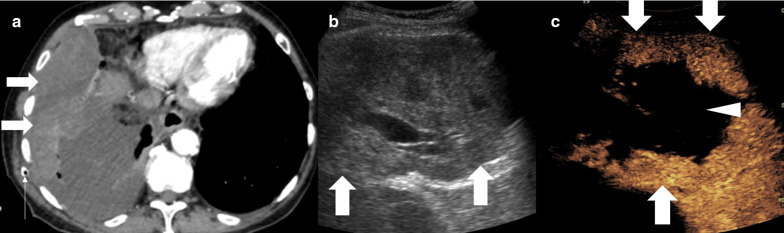
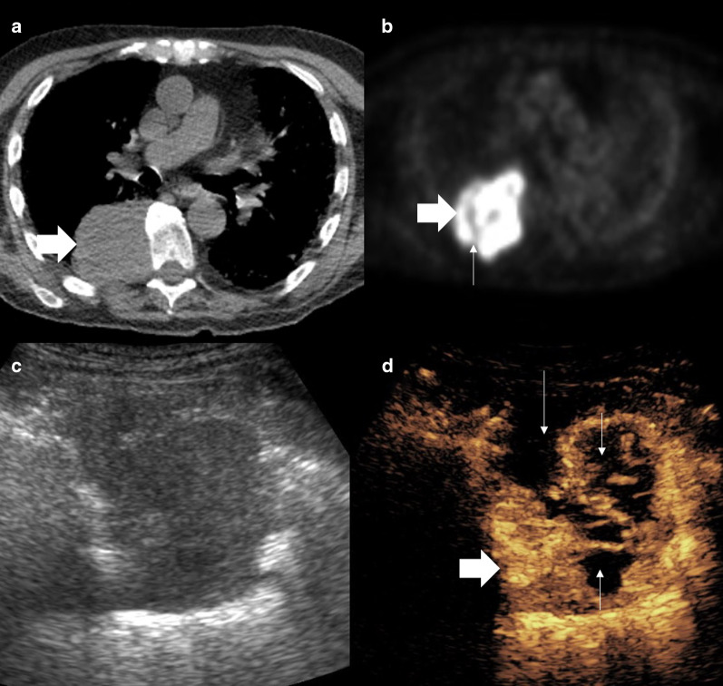
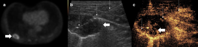
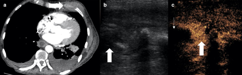
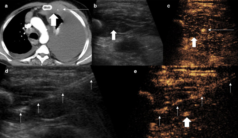
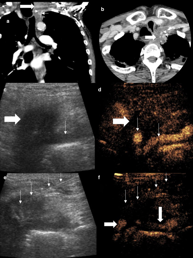
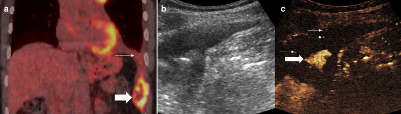
Lung cancer is one of the commonest malignancies worldwide and necessitates both early and personalised treatment. A key requirement is histological sampling with immunohistochemistry obtained usually from percutaneous biopsy. Conventionally thoracic biopsies are performed using CT guidance, but more recently, there has been development of physician led ultrasound biopsy for pleural lesions. Contrast-enhanced ultrasound (CEUS) has been increasingly used in interventional procedures and is able to offer benefits for thoracic biopsies including improving lesional visualisation and characterisation, targeting viable tissue and avoiding critical vascular structures as well as evaluating for the presence of post-procedural complications. This educational review aims to benefits of the role of CEUS in thoracic biopsies.

Keywords: Biopsy; CEUS; Contrast-enhanced ultrasound; Lung cancer; Pleural.


Conflict of interest statement

GTY has received honoraria for Siemens and Bracco. PSS has received honoraria from Siemens, Samsung, Hitachi and Bracco, and consulting fees from ITREAS. DYH, DR, SBP, KS have no disclosures.

Figures

Fig. 1
Fig. 1
Patient with pleural mesothelioma. a B mode ultrasound shows a relatively ill-defined lesion (arrow), which is difficult to differentiate from the striated muscle at some points (arrowheads). b CEUS shows clear hyperenhancement of the lesion (arrow) defining the borders and necrotic component (thin arrow) from the straited muscle layer (arrowhead) allowing accurate targeting for an optimal biopsy sample
Fig. 2
Fig. 2
Male patient with persistent cough. a A CT examination showed focal anterior pleural thickening (arrow). b PET-CT examination demonstrated a small focus of avidity within the pleura thickening. c B mode ultrasound defines the area of pleural thickening but not the smaller focus of avidity seen on PET-CT (d) simultaneous CEUS showed the subpleural area to be hypoenhancing (thick arrow) with a 4-mm focus of hyperenhancement (thin arrows) suggestive of the pathological tissue. e, f Simultaneous B mode and CEUS imaging with an 18-gauge biopsy needle (thin arrows) traversed through the enhancing focus (thick arrow). Histology revealed pleural adenocarcinoma
Fig. 3
Fig. 3
Male patient with recent chest drainage catheter insertion. a A CT examination showed focal heterogenous density (arrow) within a pleural effusion, thought to be a haematoma secondary to the drainage catheter insertion (thin arrow). b B mode ultrasound of the lesion (arrow) is of a predominantly solid appearance. c CEUS defines the large avascular necrotic central component (arrowhead) and a thick enhancing periphery (arrow), indicating that the biopsy had to be taken from the periphery. Histology revealed sarcomatoid mesothelioma
Fig. 4
Fig. 4
Patient with squamous cell carcinoma. a CT examination shows a solid appearing lung lesion (arrow). b PET-CT suggests small areas of necrosis (thin arrow) within the avid lesion (arrow). c, d Simultaneous B mode and CEUS images show the hyperenhancing lesion (arrow) with better appreciation of the complexity and volume of necrosis (thin arrows) indicating a deep eccentric portion required targeting for biopsy (arrow)
Fig. 5
Fig. 5
Patient with squamous cell carcinoma. a PET-CT shows a peripherally avid lesion with no central avidity (arrow). b, c Simultaneous B mode and CEUS images as the biopsy is performed. B mode demonstrates a hypoechoic lesion (thick arrow) with the biopsy needle within the lesion (thin arrow). The contrast image (c) shows the biopsy (thin arrows) is taken from the deep area of enhancement and therefore viable tissue (thick arrow)
Fig. 6
Fig. 6
Patient with (a) CT examination showing an indeterminate rib lesion (arrow) and clinical sepsis. b, c Simultaneous B mode and CEUS better defined the lesion which was not well seen on B-mode ultrasound imaging (arrow) into a solid hyperenhancing component (thin arrow) and central avascular component (thick arrow). Fluid aspiration yielded pus of the fluid component and the biopsy was inflammatory indicating osteomyelitis
Fig. 7
Fig. 7
Male patient with chest wall swelling. a A CT examination showed focal soft tissue thickening (arrow) encasing the internal mammary artery (IMA) (thin arrow). b On the B mode ultrasound, the lesion (arrow) blends with striated muscle but the IMA is not seen. c CEUS defines the hypoenhancing pathological tissue (arrow) encasing the IMA (thin arrow). d, e Simultaneous B mode and CEUS imaging with an 18-gauge biopsy needle (thin arrows) advanced to the lesion avoiding the IMA (arrow). The lesion was histologically proven lymphoma
Fig. 8
Fig. 8
Patient with Pancoast adenocarcinoma of the lung. Coronal (a) and axial (b) views show soft tissue density (thick arrows) encasing the left carotid artery and subclavian artery (thin arrows). c The tumour is seen as a hypoechoic area, ill-defined on B mode ultrasound with the subclavian artery visible on the periphery (thin arrow). d Simultaneous CEUS clearly defines the hypoenhancing lesion and the previously unseen carotid artery and subclavian artery (thin arrows). e, f Simultaneous B mode ultrasound and CEUS with the biopsy needle (thin arrows) within the hypoenhancing lesion avoiding the critical arterial structures (arrow)
Fig. 9
Fig. 9
Patient with metastatic squamous cell carcinoma. a PET-CT examination shows a peripherally avid lesion in the costophrenic angle (arrow) with an associated pleural effusion (thin arrow). b, c Simultaneous B mode and CEUS images post-biopsy shows pooling of the UCA within the pleural effusion (thick arrow) which was static on dynamic imaging and single microbubbles (thin arrow) within the otherwise anechoic effusion. The findings are of post-biopsy haemorrhage which ceased without intervention

References

    1. Miranda-Filho A, Piñeros M, Bray F. The descriptive epidemiology of lung cancer and tobacco control: a global overview 2018. Salud Publica Mex. 2020;61:219–229. doi: 10.21149/10140. - DOI - PubMed
    1. Veltri A, Bargellini I, Giorgi L, Almeida PA, Akhan O. CIRSE guidelines on percutaneous needle biopsy (PNB) Cardiovasc Intervent Radiol. 2017;40(10):1501–1513. doi: 10.1007/s00270-017-1658-5. - DOI - PubMed
    1. Lee MH, Lubner MG, Hinshaw JL, Pickhardt PJ. Ultrasound guidance versus CT guidance for peripheral lung biopsy: performance according to lesion size and pleural contact. AJR Am J Roentgenol. 2018;210(3):W110–W117. doi: 10.2214/AJR.17.18014. - DOI - PubMed
    1. Hallifax RJ, Corcoran JP, Ahmed A, et al. Physician-based ultrasound-guided biopsy for diagnosing pleural disease. Chest. 2014;146(4):1001–1006. doi: 10.1378/chest.14-0299. - DOI - PubMed
    1. Sidhu PS, Cantisani V, Dietrich CF, et al. The EFSUMB guidelines and recommendations for the clinical practice of contrast-enhanced ultrasound (CEUS) in non-hepatic applications: update 2017 (long version) Ultraschall Med. 2018;39(02):e2–44. doi: 10.1055/a-0586-1107. - DOI - PubMed

LinkOut - more resources