. 2021 Jan 27;32(1):18.
doi: 10.1007/s10856-020-06473-8.

PI3K/AKT signaling drives titanium-induced angiogenic stimulus

Affiliations

PI3K/AKT signaling drives titanium-induced angiogenic stimulus

Bruna Rodrigues Martins et al. J Mater Sci Mater Med. .

Abstract

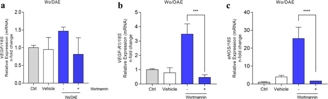
Although osseointegration and clinical success of titanium (Ti)-implanted materials depend on neovascularization in the reactional peri-implant tissue, very little has been achieved considering the Ti-molecules release on the behavior of endothelial cells. To address this issue, we challenged endothelial cells (HUVECs) with Ti-enriched medium obtained from two types of commercial titanium surfaces [presenting or not dual-acid etching (DAE)] up to 72 h to allow molecular machinery analysis. Our data show that the Ti-enriched medium provokes significant stimulus of angiogenesis-related machinery in endothelial cells by upexpressing VEGFR1, VEGFR2, VEGF, eNOS, and iNOS genes, while the PI3K/Akt signaling pathway was also significantly enhanced. As PI3K/AKT signaling was related to angiogenesis in response to vascular endothelial growth factor (VEGF), we addressed the importance of PI3K/Akt upon Ti-enriched medium responses by concomitantly treating the cells with wortmannin, a well-known PI3K inhibitor. Wortmannin suppressed the angiogenic factors, because VEGF, VEGFR1, and eNOS genes were downregulated in those cells, highlighting the importance of PI3K/AKT signaling on driving angiogenic phenotype and angiogenesis performance within the peri-implant tissue reaction. In conjunction, these data reinforce that titanium-implantable devices modify the metabolism of surrounding cells, such as endothelial cells, probably coupling osteogenesis and angiogenesis processes in peri-implant tissue and then contributing to successfully osseointegration of biomedical titanium-based devices.


Conflict of interest statement

The authors declare that they have no conflict of interest.

Figures

Fig. 1
Fig. 1
Experimental design of this study. To evaluate the biological response of endothelial cells to TiO2, the Ti-enriched medium was obtained by incubating Ti-based materials in the cell culture medium (FBS free), as recommended by ISO 10993:2016 (part 5). Thereafter, the Ti-enriched medium was used to challenge endothelial cells up to 72 h, when the samples were collected for the analysis. The main focus here was to evaluate whether there is some association of survival signaling an angiogenic stimulus of titanium, which could better support comprehension about their biocompatibility and dynamic relationship with the surrounding tissue
Fig. 2
Fig. 2
Survival signaling was evaluated by measuring the phosphorylation profile of AKT. To understand whether survival signaling was required by endothelial cells responding to Ti-enriched medium (72 h), the cells were subjected to Ti-enriched medium obtained by incubating two different conditions of titanium discs [double acid-etching (DAE) treatment (W/DAE) and without DAE (Wo/DAE)] into cell culture medium up to 24 h. After subjecting the cells up to 72 h, the samples were obtained to allow the gene expression of AKT (a) and protein performance (b, c) by evaluating AKT phosphorylation. GADPH was considered a housekeeping gene. Statistics: the value obtained to Ctrl was considered 1, and the relative values obtained to W/DAE or Wo/DAE are shown in fold‐changes. One-way ANOVA with multiple comparisons were applied to compare all pairs of groups. Differences were considered significant when *p = 0.04 and ****p < 0.0001 when compared with the Ctrl; and ****p < 0.0001 when compared with W/DAE group
Fig. 3
Fig. 3
Angiogenic stimulus of titanium (Ti) on endothelial cells. The samples were collected respecting the already described methodology to allow qPCR technology performance. To address the angiocrine effect of Ti on endothelial cells, we investigated VEGFR1 (a), VEGFR2 (b), VEGF (c), eNOS (d), and iNOS (e) genes. The GADPH gene was considered the housekeeping gene and used to normalize the values. Statistics: One-way ANOVA with multiple comparisons was used to compare all pairs of groups, and the differences were considered significant when a VEGFR1 ***p = 0.0009 and **p = 0.0019; b VEGFR2 **p = 0.0014 and **p = 0.0032; c VEGF **p = 0.004 and *p = 0.02; d eNOS ***p = 0.0006 and ***p = 0.0008; and e INOS ***p = 0.0002 and ****p < 0.0001. Black (*) when compared with Ctrl and red (*) when compared with W/DAE
Fig. 4
Fig. 4
Wortmannin targeting PI3K suppresses the angiogenic stimulus of titanium (Ti). The samples were collected respecting the already described methodology to allow qPCR technology performance. To address whether PI3K/AKT is involved in endothelial responding to the Ti-enriched medium, we reanalyzed the already known performance of genes, as was shown in Fig. 3, but now treated with wortmannin, a specific chemical inhibitor of PI3K (upstream to AKT). Thus, the suppression of the angiogenic stimulus by Ti is clear since VEGF (a), VEGFR1 (b), and eNOS (c) genes were significantly down expressed when PI3K was inhibited. The 18S gene was considered the housekeeping gene and used to normalize the values. Statistics: The statistical analysis test used was one-way ANOVA with multiple comparisons to compare all pairs of groups, and the figure specifies the differences between the absence (−) and the presence (+) of the Wortmannin. Differences were considered significant when b VEGFR1 ***p = 0.0002 and c eNOS ****p < 0.0001

References

    1. Coelho PG, Granjeiro JM, Romanos GE, Suzuki M, Silva NRF, Cardaropoli G, et al. Basic research methods and current trends of dental implant surfaces. J Biomed Mater Res B Appl Biomater. 2009;88:579–96. doi: 10.1002/jbm.b.31264. - DOI - PubMed
    1. Bonfante EA, Coelho PG. A critical perspective on mechanical testing of implants and prostheses. Adv Dent Res. 2016;28:18–27. doi: 10.1177/0022034515624445. - DOI - PubMed
    1. Dohan Ehrenfest DM, Coelho PG, Kang B-S, Sul Y-T, Albrektsson T. Classification of osseointegrated implant surfaces: materials, chemistry and topography. Trends Biotechnol. 2010;28:198–206. doi: 10.1016/j.tibtech.2009.12.003. - DOI - PubMed
    1. Raines AL, Berger MB, Patel N, Hyzy SL, Boyan BD, Schwartz Z. VEGF-A regulates angiogenesis during osseointegration of Ti implants via paracrine/autocrine regulation of osteoblast response to hierarchical microstructure of the surface. J Biomed Mater Res A. 2019;107:423–33. doi: 10.1002/jbm.a.36559. - DOI - PMC - PubMed
    1. Chen W, Xu K, Tao B, Dai L, Yu Y, Mu C, et al. Multilayered coating of titanium implants promotes coupled osteogenesis and angiogenesis in vitro and in vivo. Acta Biomater. 2018;74:489–504. doi: 10.1016/j.actbio.2018.04.043. - DOI - PubMed

MeSH terms

LinkOut - more resources