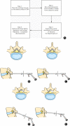
Full-Endoscopic Foraminotomy with a Novel Large Endoscopic Trephine for Severe Degenerative Lumbar Foraminal Stenosis at L5 S1 Level: An Advanced Surgical Technique
- PMID: 33506594
- PMCID: PMC7957400
- DOI: 10.1111/os.12924
Full-Endoscopic Foraminotomy with a Novel Large Endoscopic Trephine for Severe Degenerative Lumbar Foraminal Stenosis at L5 S1 Level: An Advanced Surgical Technique
Abstract
To (i) introduce the technical notes of a novel full-endoscopic foraminotomy with a large endoscopic trephine for the treatment of severe degenerative lumbar foraminal stenosis at L5 S1 level; (ii) assess the primary clinical outcomes of this technique; (iii) compare the effectiveness of this full-endoscopic foraminotomy technique and other previous techniques for lumbar foraminal stenosis. From January 2019 to August 2019, a retrospective study of L5 S1 severe degenerative lumbar foraminal stenosis was performed in our center. All patients who were diagnosed with severe foraminal stenosis at L5 S1 level and failed conservative treatment for at least 6 weeks were identified. Patients with segmental instability or other coexisting contraindications were excluded. A total of 21 patients were enrolled in the study. All patients were treated by full-endoscopic foraminotomy using large endoscopic trephine. The visual analogue scale (VAS) and Oswestry disability index (ODI) were evaluated preoperatively and at 1, 3, 6 months, and 1 year after the surgery, and the modified MacNab criteria were used to evaluate clinical outcomes at the last follow-up. There were 10 males and 11 females with a mean age of 66.38 ± 9.51 years. Five patients had a history of lumbar surgery. The mean operative time was 63.57 ± 25.74 min. The mean follow-up time was 13.29 ± 1.38 months. The mean postoperative hospital stay time was 1.29 ± 0.56 days. The mean preoperative VAS score significantly decreased from 7.38 ± 1.02 to 2.76 ± 1.09 (t = 19.759, P < 0.01), 2.25 ± 1.02 (t = 21.508, P < 0.01), 1.60 ± 1.05 (t = 31.812, P < 0.01), and 1.45 ± 1.10 (t = 25.156, P < 0.01) at 1 month, 3 months, 6 months, and 1 year after the operation. The mean preoperative ODI score significantly decreased from 64.66% ± 4.91% to 30.69% ± 4.59% (t = 33.724, P < 0.01), 29.44% ± 4.50% (t = 32.117, P < 0.01), 24.22% ± 4.14% (t = 33.951, P < 0.01), and 22.44% ± 4.94% (t = 30.241, P < 0.01) at 1 month, 3 months, 6 months, and 1 year after the operation. At the last follow-up, 19 patients (90.48%) got excellent or good outcomes. One patient suffered postoperative dysesthesia, and the symptoms were controlled by conversion treatment. One patient took revision surgery due to the incomplete decompression. There were no other major complications. Percutaneous endoscopic decompression is minimally invasive spine surgery. However, the application of endoscopic decompression for L5 S1 foraminal stenosis is relatively difficult due to the high iliac crest and narrow foramen. Full-endoscopic foraminotomy with the large endoscopic trephine is an effective and safe technique for the treatment of degenerative lumbar foraminal stenosis.
Keywords: Endoscopic trephine; Foraminal stenosis; Full-endoscopic foraminotomy; Lumbar spinal stenosis.
© 2021 The Authors. Orthopaedic Surgery published by Chinese Orthopaedic Association and John Wiley & Sons Australia, Ltd.
Figures


Similar articles
-
Uniportal Full-endoscopic Foraminotomy for Lumbar Foraminal Stenosis: Clinical Characteristics and Functional Outcomes.Orthop Surg. 2024 Aug;16(8):1861-1870. doi: 10.1111/os.14102. Epub 2024 Jun 6. Orthop Surg. 2024. PMID: 38841821 Free PMC article.
-
Effect of Dorsal Root Ganglion Retraction in Endoscopic Lumbar Decompressive Surgery for Foraminal Pathology: A Retrospective Cohort Study of Interlaminar Contralateral Endoscopic Lumbar Foraminotomy and Discectomy versus Transforaminal Endoscopic Lumbar Foraminotomy and Discectomy.World Neurosurg. 2021 Apr;148:e101-e114. doi: 10.1016/j.wneu.2020.12.176. Epub 2021 Jan 11. World Neurosurg. 2021. PMID: 33444831
-
Early experience with endoscopic revision of lumbar spinal fusions.Neurosurg Focus. 2016 Feb;40(2):E10. doi: 10.3171/2015.10.FOCUS15503. Neurosurg Focus. 2016. PMID: 26828879
-
Systematic Review of Current Literature on Clinical Outcomes of Uniportal Interlaminar Contralateral Endoscopic Lumbar Foraminotomy for Foraminal Stenosis.World Neurosurg. 2022 Dec;168:392-397. doi: 10.1016/j.wneu.2022.04.130. World Neurosurg. 2022. PMID: 36527218
-
Efficacy and Safety of Full-endoscopic Decompression via Interlaminar Approach for Central or Lateral Recess Spinal Stenosis of the Lumbar Spine: A Meta-analysis.Spine (Phila Pa 1976). 2018 Dec 15;43(24):1756-1764. doi: 10.1097/BRS.0000000000002708. Spine (Phila Pa 1976). 2018. PMID: 29794584 Review.
Cited by
-
Uniportal Full-endoscopic Foraminotomy for Lumbar Foraminal Stenosis: Clinical Characteristics and Functional Outcomes.Orthop Surg. 2024 Aug;16(8):1861-1870. doi: 10.1111/os.14102. Epub 2024 Jun 6. Orthop Surg. 2024. PMID: 38841821 Free PMC article.
-
One-Stage Percutaneous Endoscopic Lumbar Discectomy for Symptomatic Double-Level Contiguous Adolescent Lumbar Disc Herniation.Orthop Surg. 2021 Jul;13(5):1532-1539. doi: 10.1111/os.13097. Epub 2021 Jun 3. Orthop Surg. 2021. PMID: 34080296 Free PMC article.
-
Clinical Application of Large Channel Endoscopic Systems with Full Endoscopic Visualization Technique in Lumbar Central Spinal Stenosis: A Retrospective Cohort Study.Pain Ther. 2022 Dec;11(4):1309-1326. doi: 10.1007/s40122-022-00428-3. Epub 2022 Sep 3. Pain Ther. 2022. PMID: 36057015 Free PMC article.
-
Complications and Management of Endoscopic Spinal Surgery.Neurospine. 2023 Mar;20(1):56-77. doi: 10.14245/ns.2346226.113. Epub 2023 Mar 31. Neurospine. 2023. PMID: 37016854 Free PMC article.
-
[Research progress of different minimally invasive spinal decompression in lumbar spinal stenosis].Zhongguo Xiu Fu Chong Jian Wai Ke Za Zhi. 2023 Jul 15;37(7):895-900. doi: 10.7507/1002-1892.202303110. Zhongguo Xiu Fu Chong Jian Wai Ke Za Zhi. 2023. PMID: 37460188 Free PMC article. Review. Chinese.
References
-
- Yuan SG, Wen YL, Zhang P, Li YK. Ligament, nerve, and blood vessel anatomy of the lateral zone of the lumbar intervertebral foramina. Int Orthop, 2015, 39: 2135–2141. - PubMed
-
- Hasue M, Kunogi J, Konno S, Kikuchi S. Classification by position of dorsal root ganglia in the lumbosacral region. Spine (Phila Pa 1976), 1989, 14: 1261–1264. - PubMed
-
- Hasegawa T, An HS, Haughton VM, Nowicki BH. Lumbar foraminal stenosis: critical heights of the intervertebral discs and foramina. A cryomicrotome study in cadavera. J Bone Joint Surg Am, 1995, 77: 32–38. - PubMed
-
- Jenis LG, An HS. Spine update. Lumbar foraminal stenosis. Spine (Phila Pa 1976), 2000, 25: 389–394. - PubMed
-
- Akhgar J, Terai H, Rahmani MS, et al. Anatomical analysis of the relation between human ligamentum flavum and posterior spinal bony prominence. J Orthop Sci, 2017, 22: 260–265. - PubMed
Publication types
MeSH terms
Grants and funding
LinkOut - more resources
Full Text Sources
Other Literature Sources
Medical

