Foramen magnum meningiomas: a systematic review and meta-analysis
- PMID: 33507444
- PMCID: PMC8490226
- DOI: 10.1007/s10143-021-01478-5
Foramen magnum meningiomas: a systematic review and meta-analysis
Abstract
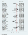
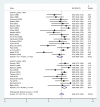
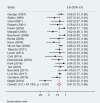
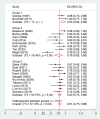
Foramen magnum meningiomas (FMMs) account for 1.8-3.2% of all meningiomas. With this systematic review and meta-analysis, our goal is to detail epidemiology, clinical features, surgical aspects, and outcomes of this rare pathology. Using PRISMA 2015 guidelines, we reviewed case series, mixed series, or retrospective observational cohorts with description of surgical technique, patient and lesion characteristics, and pre- and postoperative clinical status. A meta-analysis was performed to search for correlations between meningioma characteristics and rate of gross total resection (GTR). We considered 33 retrospective studies or case series, including 1053 patients, mostly females (53.8%), with a mean age of 52 years. The mean follow-up was of 51 months (range 0-258 months). 65.6% of meningiomas were anterior, and the mean diameter was of 29 mm, treated with different surgical approaches. Postoperatively, 17.2% suffered complications (both surgery- and non-surgery-related) and 2.5% had a recurrence. The Karnofsky performance score improved in average after surgical treatment (75 vs. 81, p < 0.001). Our meta-analysis shows significant rates of GTR in cohorts with a majority of posterior and laterally located FMM (p = 0.025) and with a mean tumor less than 25 mm (p < 0.05). FMM is a rare and challenging pathology whose treatment should be multidisciplinary, focusing on quality of life. Surgery still remains the gold standard and aim at maximal resection with neurological function preservation. Adjuvant therapies are needed in case of subtotal removal, non-grade I lesions, or recurrence. Specific risk factors for recurrence, other than Simpson grading, need further research.
Keywords: Classification; Foramen magnum; Meningioma; Meta-analysis; Outcome; Surgery; Systematic review.
© 2021. The Author(s).
Conflict of interest statement
The authors declare no competing interests.
Figures


References
Publication types
MeSH terms
LinkOut - more resources
Full Text Sources
Other Literature Sources

