. 2021 Feb 10;13(5):6011-6022.
doi: 10.1021/acsami.0c20607. Epub 2021 Jan 28.

Lipid-Polyglutamate Nanoparticle Vaccine Platform

Affiliations

Lipid-Polyglutamate Nanoparticle Vaccine Platform

Dorien Van Lysebetten et al. ACS Appl Mater Interfaces. .

Abstract

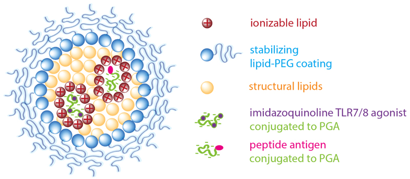
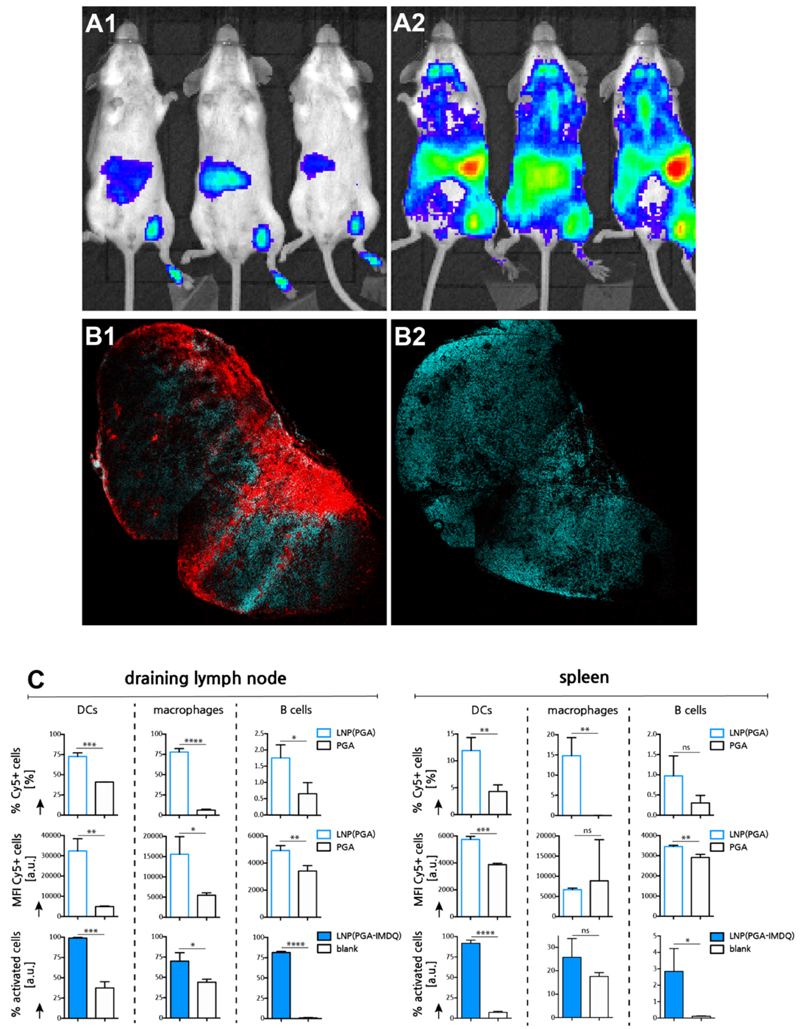
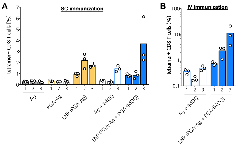
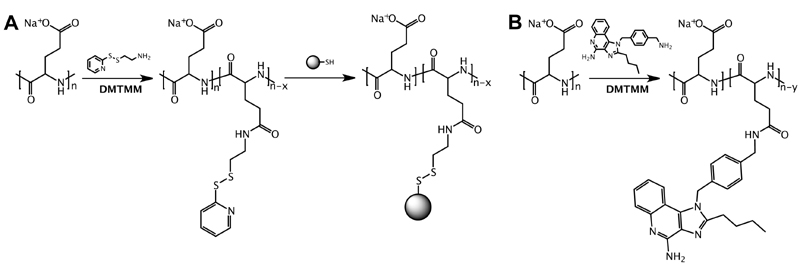
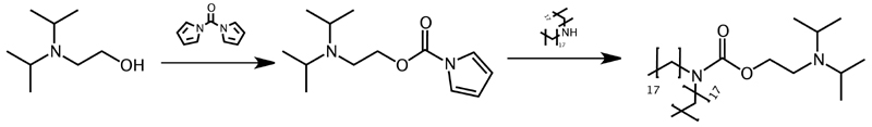
Peptide-based subunit vaccines are attractive in view of personalized cancer vaccination with neo-antigens, as well as for the design of the newest generation of vaccines against infectious diseases. Key to mounting robust antigen-specific immunity is delivery of antigen to antigen-presenting (innate immune) cells in lymphoid tissue with concomitant innate immune activation to promote antigen presentation to T cells and to shape the amplitude and nature of the immune response. Nanoparticles that co-deliver both peptide antigen and molecular adjuvants are well suited for this task. However, in the context of peptide-based antigen, an unmet need exists for a generic strategy that allows for co-encapsulation of peptide and molecular adjuvants due to the stark variation in physicochemical properties based on the amino acid sequence of the peptide. These properties also strongly differ from those of many molecular adjuvants. Here, we devise a lipid nanoparticle (LNP) platform that addresses these issues. Key in our concept is poly(l-glutamic acid) (PGA), which serves as a hydrophilic backbone for conjugation of, respectively, peptide antigen (Ag) and an imidazoquinoline (IMDQ) TLR7/8 agonist as a molecular adjuvant. Making use of the PGA's polyanionic nature, we condensate PGA-Ag and PGA-IMDQ into LNP by electrostatic interaction with an ionizable lipid. We show in vitro and in vivo in mouse models that LNP encapsulation favors uptake by innate immune cells in lymphoid tissue and promotes the induction of Ag-specific T cells responses both after subcutaneous and intravenous administration.

Keywords: TLR agonists; lipid nanoparticles; peptides; vaccine.


Figures

Figure 1
Figure 1. Design of lipid-polyglutamate vaccine nanoparticles.
Electrostatic interaction between carboxylic acid moieties of poly-L-glutamic acid (PGA) and cationic moieties of an ionizable lipid drive nanoparticle formation aided by hydrophobic interaction. A PEG-lipid provides colloidal stabilization. Antigen (Ag) and an imidazoquinoline TLR7/8 agonist (IMDQ) are separately conjugated to PGA.
Figure 2
Figure 2. (A) Size distribution curves measured by DLS of LNP formulations listed in Table 1.
Figure 3
Figure 3. Physicochemical characterization of LNP.
(A) Asymmetric field flow fractionation (AFFF) analysis of LNP. Panel A1 depicts multiple parameters as function of the elution time, panel A2 depicts the calculated size distribution based on the light scattering signal. (B) AFFF analysis used to determine encapsulation of OG-PGA in LNP. Panel B1 depicts the fluorescence signal in the time interval where OG-PGA elutes. Panel B2 depicts the light scattering signal in the time interval where the LNP elute. (C) LNP analysis by fluorescence correlation spectroscopy (FCS). Panel C1 depicts normalized autocorrelation curves (evaluated from measuring fluorescence fluctuations in the OG(-PGA) signal) and their fits (solid lines) that yielded the radii of PGA and LNP in aqueous medium. Panel C2 depicts normalized autocorrelation curves (calculated from measuring fluorescence fluctuations in the OG(-PGA) signal) and the corresponding fit (solid line) yielding the radius of LNP in human blood plasma. Panel C3 depicts time fluorescence fluctuations (time traces) of the OG(-PGA) and Cy5(-DOPE) signals. Panel C4 depicts auto- and cross-correlation curves the OG(-PGA) and Cy5(-DOPE) signals from LNP in aqueous medium.
Figure 4
Figure 4. In vitro immuno-biological characterization of LNP.
(A) Cell viability measured by MTT assay. (n=6) (B) Flow cytometry analysis if DC2.4 cells pulsed with single-dye or double-dye labeled LNP. (C) Confocal microscopy of DC2.4 cells pulsed with LNP. Scale bar represents 15 microns. (D) Dose-response innate activation measured by the RAW Blue reporter cell assay (n=3). (E) Flow cytometry analysis of DC2.4 cells pulsed with different concentration of antigen loaded LNP followed by immunostaining with the OVA-Kb antibody (n=3; t-test *: p<0.05).
Figure 5
Figure 5. In vivo lymphatic transportation and innate immune activation by LNP.
(A) Bioluminescence imaging of IFNb-luciferase reporter mouse 4h post injection. (A1: PGA-IMDQ; A2: LNP(PGA-IMDQ)) (B) Confoca microscopy image of a sectioned popliteal lymph node, 24h post injection of LNP in the foot pad. (cyan: DAPI, red:Cy5-PGA loaded LNP). Note that some irregularities in the image are due to stitching. (B1 : LNP(Cy5-PGA); B2: Cy5-PGA) (C) Flow cytometry analysis of LNP uptake by innate immune cells and innate immune cell activation in the draining inguinal lymph node) 24h post injection of LNP in the tail base (n=3; t-test *: p<0.05, **:p<0.01, ***:p<0.001, ****:p<0.0001).
Figure 6
Figure 6. In vivo adaptive immune response.
(A) Subcutaneous immunization in tail base. (n=3) (B) Intravenous immunization in tail vain. (n=3)
Scheme 1
Scheme 1. Poly-L-glutamic acid (PGA) conjugation.
(A) For conjugation of peptide antigen (Ag), PGA is first functionalized with a pyridyldisulfide moiety followed by conjugation of the Ag to a terminal cysteine residue via a disulfide bond. (B) For conjugation of the imidazoquinoline TLR7/8 agonist IMDQ, aliphatic amine of IMDQ is conjugated to a carboxylic acid moiety of PGA through an amide bond.
Scheme 2
Scheme 2. Synthesis of the ionizable lipid DIPADS through carbamate conjugation of distearylamine to 2-(diisopropylamino)ethanol.

References

    1. Melief CJM, van Hall T, Arens R, Ossendorp F, van der Burg SH. Therapeutic Cancer Vaccines. J Clin Invest. 2015;125(9):1–12. - PMC - PubMed
    1. Mancini RJ, Stutts L, Ryu KA, Tom JK, Esser-Kahn AP. Directing the Immune System with Chemical Compounds. ACSChem Biol. 2014;9(5):1075–1085. - PMC - PubMed
    1. Schumacher TN, Schreiber RD. Neoantigens in Cancer Immunotherapy. Science (80-. ) 2015;348(6230):69–74. - PubMed
    1. Irvine DJ, Hanson MC, Rakhra K, Tokatlian T. Synthetic Nanoparticles for Vaccines and Immunotherapy. Chem Rev. 2015;115(19):11109–11146. - PMC - PubMed
    1. Moon JJ, Suh H, Bershteyn A, Stephan MT, Liu H, Huang B, Sohail M, Luo S, Ho Um S, Khant H, Goodwin JT, et al. Interbilayer-Crosslinked Multilamellar Vesicles as Synthetic Vaccines for Potent Humoral and Cellular Immune Responses. Nat Mater. 2011;10(3):243–251. - PMC - PubMed