Prostate cancer GTV delineation with biparametric MRI and 68Ga-PSMA-PET: comparison of expert contours and semi-automated methods
- PMID: 33507812
- PMCID: PMC8011238
- DOI: 10.1259/bjr.20201174
Prostate cancer GTV delineation with biparametric MRI and 68Ga-PSMA-PET: comparison of expert contours and semi-automated methods
Abstract
Objective: The optimal method for delineation of dominant intraprostatic lesions (DIL) for targeted radiotherapy dose escalation is unclear. This study evaluated interobserver and intermodality variability of delineations on biparametric MRI (bpMRI), consisting of T2 weighted (T2W) and diffusion-weighted (DWI) sequences, and 68Ga-PSMA-PET/CT; and compared manually delineated GTV contours with semi-automated segmentations based on quantitative thresholding of intraprostatic apparent diffusion coefficient (ADC) and standardised uptake values (SUV).
Methods: 16 patients who had bpMRI and PSMA-PET scanning performed prior to any treatment were eligible for inclusion. Four observers (two radiation oncologists, two radiologists) manually delineated the DIL on: (1) bpMRI (GTVMRI), (2) PSMA-PET (GTVPSMA) and (3) co-registered bpMRI/PSMA-PET (GTVFused) in separate sittings. Interobserver, intermodality and semi-automated comparisons were evaluated against consensus Simultaneous Truth and Performance Level Estimation (STAPLE) volumes, created from the relevant manual delineations of all observers with equal weighting. Comparisons included the Dice Similarity Coefficient (DSC), mean distance to agreement (MDA) and other metrics.
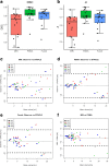
Results: Interobserver agreement was significantly higher (p < 0.05) for GTVPSMA (DSC: 0.822, MDA: 1.12 mm) and GTVFused (DSC: 0.787, MDA: 1.34 mm) than for GTVMRI (DSC: 0.705, MDA 2.44 mm). Intermodality agreement between GTVMRI and GTVPSMA was low (DSC: 0.440, MDA: 4.64 mm). Agreement between semi-automated volumes and consensus GTV was low for MRI (DSC: 0.370, MDA: 8.16 mm) and significantly higher for PSMA-PET (0.571, MDA: 4.45 mm, p < 0.05).
Conclusion: 68Ga-PSMA-PET appears to improve interobserver consistency of DIL localisation vs bpMRI and may be more viable for simple quantitative delineation approaches; however, more sophisticated approaches to semi-automatic delineation factoring for patient- and disease-related heterogeneity are likely required.
Advances in knowledge: This is the first study to evaluate the interobserver variability of prostate GTV delineations with co-registered bpMRI and 68Ga-PSMA-PET.
Figures


References
-
- Lips IM, van der Heide UA, Haustermans K, van Lin ENJT, Pos F, Franken SPG, et al. . Single blind randomized phase III trial to investigate the benefit of a focal lesion ablative microboost in prostate cancer (FLAME-trial): study protocol for a randomized controlled trial. Trials 2011; 12: 255. doi: 10.1186/1745-6215-12-255 - DOI - PMC - PubMed
-
- Draulans C, De Roover R, van der Heide UA, Haustermans K, Pos F, Smeenk RJ, et al. . Stereotactic body radiation therapy with optional focal lesion ablative microboost in prostate cancer: topical review and multicenter consensus. Radiother Oncol 2019; 140: 131–42. doi: 10.1016/j.radonc.2019.06.023 - DOI - PubMed
-
- Stoyanova R, Chinea F, Kwon D, Reis IM, Tschudi Y, Parra NA, et al. . An Automated Multiparametric MRI Quantitative Imaging Prostate Habitat Risk Scoring System for Defining External Beam Radiation Therapy Boost Volumes. Int J Radiat Oncol Biol Phys 2018; 102: 821–9. doi: 10.1016/j.ijrobp.2018.06.003 - DOI - PMC - PubMed
Publication types
MeSH terms
Substances
LinkOut - more resources
Full Text Sources
Other Literature Sources
Medical
Miscellaneous

