. 2021 Mar 15;131(6):e143861.
doi: 10.1172/JCI143861.

An abundant biliary metabolite derived from dietary omega-3 polyunsaturated fatty acids regulates triglycerides

Affiliations

An abundant biliary metabolite derived from dietary omega-3 polyunsaturated fatty acids regulates triglycerides

Trisha J Grevengoed et al. J Clin Invest. .

Abstract

Omega-3 fatty acids from fish oil reduce triglyceride levels in mammals, yet the mechanisms underlying this effect have not been fully clarified, despite the clinical use of omega-3 ethyl esters to treat severe hypertriglyceridemia and reduce cardiovascular disease risk in humans. Here, we identified in bile a class of hypotriglyceridemic omega-3 fatty acid-derived N-acyl taurines (NATs) that, after dietary omega-3 fatty acid supplementation, increased to concentrations similar to those of steroidal bile acids. The biliary docosahexaenoic acid-containing (DHA-containing) NAT C22:6 NAT was increased in human and mouse plasma after dietary omega-3 fatty acid supplementation and potently inhibited intestinal triacylglycerol hydrolysis and lipid absorption. Supporting this observation, genetic elevation of endogenous NAT levels in mice impaired lipid absorption, whereas selective augmentation of C22:6 NAT levels protected against hypertriglyceridemia and fatty liver. When administered pharmacologically, C22:6 NAT accumulated in bile and reduced high-fat diet-induced, but not sucrose-induced, hepatic lipid accumulation in mice, suggesting that C22:6 NAT is a negative feedback mediator that limits excess intestinal lipid absorption. Thus, biliary omega-3 NATs may contribute to the hypotriglyceridemic mechanism of action of fish oil and could influence the design of more potent omega-3 fatty acid-based therapeutics.

Keywords: Endocrinology; Fatty acid oxidation; Gastroenterology; Lipoproteins.


Conflict of interest statement

Conflict of interest: The authors have declared that no conflict of interest exists.

Figures

Figure 1
Figure 1. NATs are enriched in bile, and omega-3 fatty acid supplementation increases C22:6 NAT amounts.
(A) Representative chromatogram of murine bile after 3 days of a lard-based HFD or a fish oil diet. The indicated peaks denote steroidal bile acids or C22:6 and C22:7 NATs. Insets show the structures of the taurine-conjugated biliary lipids taurocholate and C22:6-NAT. Biliary (B) and plasma (C) NATs after 3 days of HFD or fish oil feeding (n = 8–10). (D) Human plasma after EPA/DHA supplementation (13.1 g/week) or placebo oil (n = 14). Data are presented as the mean ± SEM. *P < 0.05 between treatments by FDR-corrected t test (AC) or 2-way ANOVA (D).
Figure 2
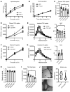
Figure 2. C22:6 NAT lowers lipid absorption by inhibiting TAG degradation.
(A and B) Plasma TAG with gavage of olive oil (lipid absorption; n = 11–12) or after inhibition of lipoprotein lipase alone (VLDL secretion; n = 4). (CE) 3H in plasma or small intestine after lipoprotein lipase inhibition and oral gavage of oil with [3H]triolein ± 100 μM NAT (n = 4–7). (F and G) 14C in plasma or small intestine after lipoprotein lipase inhibition and oral gavage of oil with [14C]oleic acid ± 100 μM C22:6 NAT (n = 7–8). (H) Lipase activity measured using 4-nitrophenyl palmitate ± 100 μM NAT (n = 2 separate experiments in triplicate). (I) Lipase-mediated TAG hydrolysis measured using [3H]triolein in a taurocholate emulsion ± 100 μM NAT (n = 3 separate experiments in triplicate). (J) Representative images and quantification of size of bovine bile micelles ± 500 μM C22:6 NAT imaged by cryo-EM. Micelles are the small, dark circles. n = 96 (vehicle); n = 772 (C22:6 NAT). Scale bars: 200 nm. Data are presented as the mean ± SEM. *P < 0.05 between treatments using t tests (A, B, D, F, and G) or 1-way ANOVA (C, E, H, and I) and ****P < 0.001 between conditions using a Mann-Whitney U test (J). FA, fatty acid.
Figure 3
Figure 3. Genetic ablation of NAT hydrolysis impairs lipid absorption.
(A) Biliary NAT in FAAH-WT and FAAH-S268D mice (n = 6–8). (B and C) Plasma TAG after gavage of sunflower oil (lipid absorption; n = 7–8) or after inhibition of lipoprotein lipase alone (VLDL secretion; n = 7). (D) Fecal acylglycerol after 4 weeks of very HFD feeding (60% kcal from fat; n = 6–8). (E) Oral maltodextrin digestion (n = 8). (F) Plasma FITC-dextran 4 hours after oral gavage (n = 5–6). (G) Upper gut transit measured 30 minutes after gavage of a mixed meal containing 10% charcoal (n = 8). (H) Hepatic bile secretion rate (n = 6–8). (I) Biliary steroidal bile acids (n = 6–8). TCA, taurocholic acid; TCDCA, taurochenodeoxycholic acid; TDCA, taurodeoxycholic acid; TαMCA, tauro-α-muricholic acid; TαMCA, tauro-β-muricholic acid; TUDCA, tauroursodeoxycholic acid; CA, cholic acid; GCA, glycocholic acid; ω-MCA, ω-muricholic acid; β-MCA, β-muricholic acid. (J) Hepatic gene expression (n = 5–7). Data are presented as the mean ± SEM. *P < 0.05 compared with FAAH-WT using t tests.
Figure 4
Figure 4. Impaired NAT hydrolysis alters whole-body energetics.
(A) Body composition in chow-fed, 10- to 12-week-old male mice, measured by MRI (n = 12–15). (BG) Metabolic chambers measured food intake, physical activity, heat output, respiratory exchange ratio (RER), VO2, and VCO2 over at least 72 hours (n = 6–10). Data are presented as the mean ± SEM. *P < 0.05 compared with FAAH-WT by t test.
Figure 5
Figure 5. Fish oil feeding of FAAH-S268D mice prevents hypertriglyceridemia and hepatic lipid accumulation.
Plasma (A) and biliary (B) C22:6 NAT after 3 days of a lard-based HFD (45% kcal fat) or a matched diet containing 20% kcal from fish oil (n = 8–11). (C and D) Plasma TAG with gavage of olive oil (lipid absorption; n = 9–13) or after inhibition of lipoprotein lipase alone (VLDL secretion; n = 3–4). (E) BW change over the 6-week diet period (n = 9–14). (F) Total fat and lean mass after 5 weeks of the indicated diets (n = 9–14). (G) Gonadal white adipose tissue (GWAT) weight (n = 9–14). (HJ) Plasma lipid levels (n = 9–14). (K) Liver weight (n = 9–14). (L) Liver TAG levels after 6 weeks on the indicated diets (n = 9–14). (M) Hepatic expression of lipid metabolism genes and markers of inflammation (n = 6). Data are presented as the mean ± SEM. *P < 0.05 compared with FAAH-WT mice, by 2-way ANOVA; #P < 0.05 compared with HFD feeding within the genotype, by 2-way ANOVA.
Figure 6
Figure 6. C22:6 NAT treatment of FAAH-S268D mice mimics the effects of fish oil feeding.
C22:6 NAT (10 mg/kg) was administered s.c. to FAAH-S268D mice, and plasma (A) and bile (B) were analyzed for changes in NATs (n = 3–5). (C) Plasma TAG levels with gavage of olive oil and lipoprotein lipase inhibition 2 hours after s.c. injection of C22:6 NAT (10 mg/kg) or vehicle (PBS) (n = 9–10) into FAAH-S268D mice. (DM) FAAH-S268D mice were fed a lard-based HFD (45% kcal fat) for 2 weeks and given daily s.c. C22:6 NAT (10 mg/kg) or vehicle (PBS) (n = 11–16). (D) BW change over a 2-week period. (E and F) Adipose depot weights. IWAT, inguinal white adipose tissue. (GK) Plasma values. (L) Liver TAG. (M) Histologic analysis of liver. Original magnification, ×20; scale bars: 200 μm. For lipids, the scores were defined as follows: 0: <2%; 1: 2%–5%; 2: >5% (n = 6). Data are presented as the mean ± SEM. *P < 0.05 compared with vehicle, by t test.
Figure 7
Figure 7. A HSD prevents C22:6 NAT treatment protection against fatty liver.
(AD) FAAH-S268D mice were fed a HSD for 2 weeks and received daily s.c. treatment with C22:6 NAT (10 mg/kg) or vehicle (PBS) (n = 9–10). (A) BW change. (B) GWAT weight. (C) Food intake over the last 7 days of the diet (n = 5–6). (D) Plasma values. (E) Liver TAG levels. (FI) FAAH-S268D mice fasted for 12 hours were given a s.c. injection of C22:6 NAT (10 mg/kg) or vehicle and, 2 hours later, were dosed orally with glucose or a high-fat mixed meal. Blood was collected 30 minutes after gavage (n = 9–10). Data are presented as the mean ± SEM. *P < 0.05 compared with vehicle, by t test.

Comment in

References

    1. Calder PC. Omega-3 polyunsaturated fatty acids and inflammatory processes: nutrition or pharmacology? Br J Clin Pharmacol. 2013;75(3):645–662. doi: 10.1111/j.1365-2125.2012.04374.x. - DOI - PMC - PubMed
    1. Yan JH, et al. Omega-3 polyunsaturated fatty acid supplementation and non-alcoholic fatty liver disease: a meta-analysis of randomized controlled trials. Medicine (Baltimore) 2018;97(37):e12271. doi: 10.1097/MD.0000000000012271. - DOI - PMC - PubMed
    1. Musa-Veloso K, et al. Systematic review and meta-analysis of controlled intervention studies on the effectiveness of long-chain omega-3 fatty acids in patients with nonalcoholic fatty liver disease. Nutr Rev. 2018;76(8):581–602. doi: 10.1093/nutrit/nuy022. - DOI - PMC - PubMed
    1. Skulas-Ray AC, et al. Omega-3 fatty acids for the management of hypertriglyceridemia: a science advisory from the american heart association. Circulation. 2019;140(12):E673–E691. - PubMed
    1. Bhatt DL, et al. Cardiovascular risk reduction with icosapent ethyl for hypertriglyceridemia. N Engl J Med. 2019;380(1):11–22. doi: 10.1056/NEJMoa1812792. - DOI - PubMed

Publication types

MeSH terms