. 2021 Jan 28;17(1):e1009292.
doi: 10.1371/journal.ppat.1009292. eCollection 2021 Jan.

Single cell resolution of SARS-CoV-2 tropism, antiviral responses, and susceptibility to therapies in primary human airway epithelium

Affiliations

Single cell resolution of SARS-CoV-2 tropism, antiviral responses, and susceptibility to therapies in primary human airway epithelium

Jessica K Fiege et al. PLoS Pathog. .

Abstract

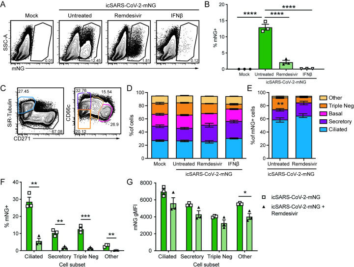
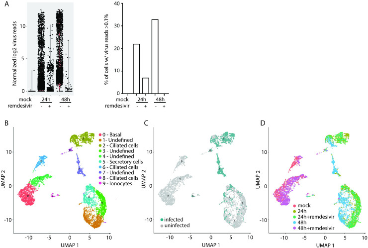
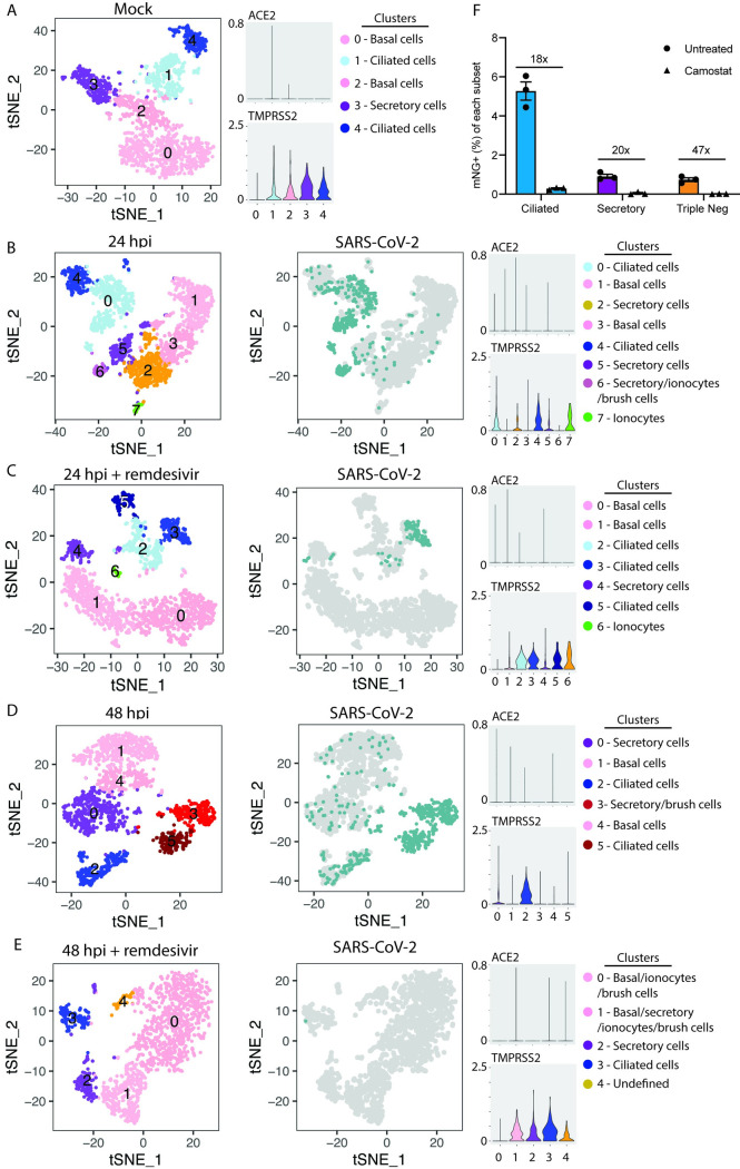
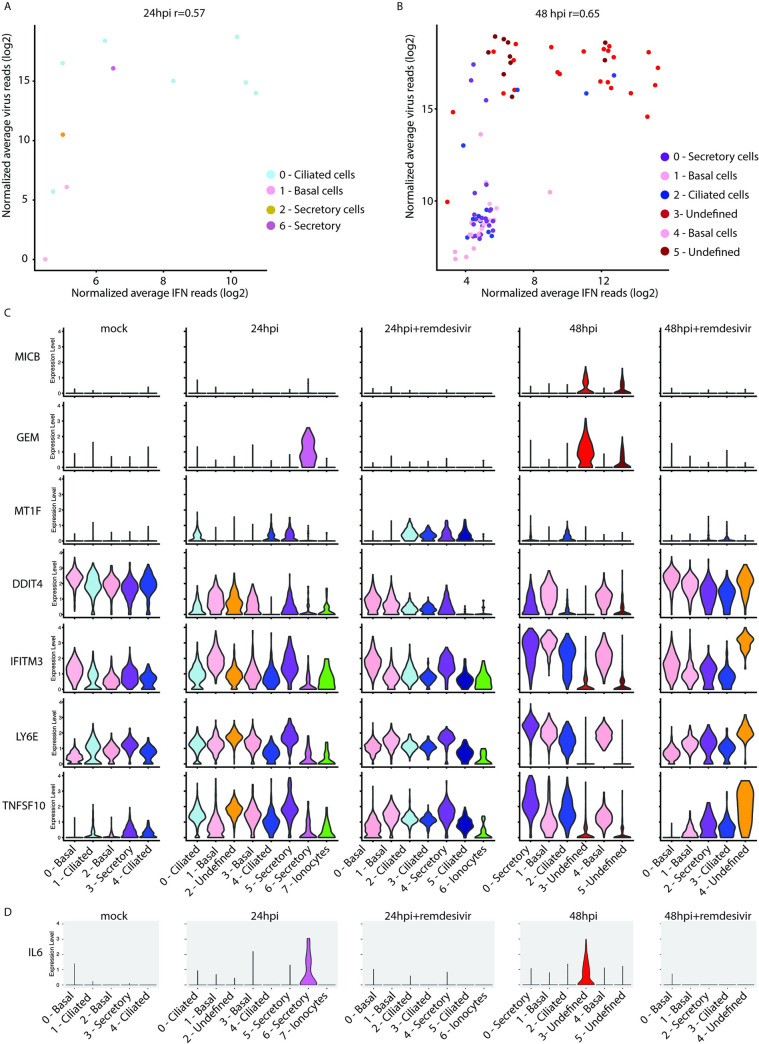
The human airway epithelium is the initial site of SARS-CoV-2 infection. We used flow cytometry and single cell RNA-sequencing to understand how the heterogeneity of this diverse cell population contributes to elements of viral tropism and pathogenesis, antiviral immunity, and treatment response to remdesivir. We found that, while a variety of epithelial cell types are susceptible to infection, ciliated cells are the predominant cell target of SARS-CoV-2. The host protease TMPRSS2 was required for infection of these cells. Importantly, remdesivir treatment effectively inhibited viral replication across cell types, and blunted hyperinflammatory responses. Induction of interferon responses within infected cells was rare and there was significant heterogeneity in the antiviral gene signatures, varying with the burden of infection in each cell. We also found that heavily infected secretory cells expressed abundant IL-6, a potential mediator of COVID-19 pathogenesis.


Conflict of interest statement

The authors have declared that no competing interests exist

Figures

Fig 1
Fig 1. SARS-CoV-2 infects multiple cell types and infection is controlled by remdesivir in primary nHTBE cells.
nHTBE cells differentiated at air-liquid interface were untreated or treated with IFN-β or remdesivir prior to infection with icSARS-CoV-2-mNG at a MOI of 2.5. Cells were analyzed 48 hpi by flow cytometry. (A) Representative plots of live cells (B) Frequency of infected cells (mNG+). (C) Representative plots of cell subset gating. Ciliated (SiR-Tubulin+ CD271-), secretory (SiR-Tubulin- CD271- CD66c+), basal (SiR-Tubulin- CD271+ CD66c-), Triple negative (SiR-Tubulin- CD271- CD66c-) and other cells (SiR-Tubulin- CD217int CD66cint) (D) Frequency of cell subsets from live cells. (E) Frequency of cell subsets of infected cells (mNG+). (F) Total frequency of mNG+ infected cell types. (G) mNG gMFI within each cell subset. The data (B, D-F) are 3 biological replicates per group +/-SEM, 1 of 2 independent experiments.
Fig 2
Fig 2. Single cell resolution of SARS-CoV-2 infection.
(A) Total viral reads per cell in each condition (left) and frequency of cells expressing at least 0.01% virus reads (right). (B) Cells from all samples were combined and analyzed by UMAP. (C) Cells with greater than 0.1% of total reads mapping to SARS-CoV-2. (D) Cells colored according to experimental condition.
Fig 3
Fig 3. Single cell resolution of SARS-CoV-2 tropism.
tSNE plots demonstrating clustering (left) and infected cells (right, blue) and volume plots for ACE2 and TMPRSS2 in mock (A), 24 hpi (B), 24 hpi with remdesivir (C), 48 hpi (D), and 48 hpi with remdesivir (E). (F) nHTBE cells were pretreated with vehicle or camostat, then infected with icSARS-CoV-2-mNG at MOI = 2.5 and analyzed for mNG in ciliated (SiR-tubulin+ CD271-), secretory cells (CD66c+ SiR-tubulin- CD271-), and triple negative cells. Numbers indicate fold change between vehicle and camostat treated cells.
Fig 4
Fig 4. Distinct antiviral responses during SARS-CoV-2 infection.
Cells from Fig 3 were analyzed at 24hpi (A) and 48hpi (B) cells expressing interferon (IFNB1, IFNL1, IFNL2, IFNL3, IFNL4) were assessed for levels of combined IFN transcripts plotted against combined SARS-CoV-2 reads. Colors indicate clusters from Fig 3B and 3D, respectively. (C) Volume plots of cells from Fig 3 for ISGs with statistically significant expression between samples. (D) Volume plot of IL-6 expression.

Update of

Similar articles

Cited by

References

    1. de Jong PM, van Sterkenburg MA, Kempenaar JA, Dijkman JH, Ponec M. Serial culturing of human bronchial epithelial cells derived from biopsies. In Vitro Cell Dev Biol Anim. 1993;29A(5):379–87. 10.1007/BF02633985 . - DOI - PubMed
    1. de Jong PM, van Sterkenburg MA, Hesseling SC, Kempenaar JA, Mulder AA, Mommaas AM, et al. Ciliogenesis in human bronchial epithelial cells cultured at the air-liquid interface. Am J Respir Cell Mol Biol. 1994;10(3):271–7. 10.1165/ajrcmb.10.3.8117445 . - DOI - PubMed
    1. Sungnak W, Huang N, Becavin C, Berg M, Queen R, Litvinukova M, et al. SARS-CoV-2 entry factors are highly expressed in nasal epithelial cells together with innate immune genes. Nat Med. 2020;26(5):681–7. 10.1038/s41591-020-0868-6 . - DOI - PMC - PubMed
    1. Jia HP, Look DC, Shi L, Hickey M, Pewe L, Netland J, et al. ACE2 receptor expression and severe acute respiratory syndrome coronavirus infection depend on differentiation of human airway epithelia. Journal of virology. 2005;79(23):14614–21. 10.1128/JVI.79.23.14614-14621.2005 - DOI - PMC - PubMed
    1. Hoffmann M, Kleine-Weber H, Schroeder S, Kruger N, Herrler T, Erichsen S, et al. SARS-CoV-2 Cell Entry Depends on ACE2 and TMPRSS2 and Is Blocked by a Clinically Proven Protease Inhibitor. Cell. 2020;181(2):271–80 e8. 10.1016/j.cell.2020.02.052 - DOI - PMC - PubMed

Publication types

MeSH terms