. 2021 Jan 28;16(1):e0245685.
doi: 10.1371/journal.pone.0245685. eCollection 2021.

Brain organoid formation on decellularized porcine brain ECM hydrogels

Affiliations

Brain organoid formation on decellularized porcine brain ECM hydrogels

Robin Simsa et al. PLoS One. .

Abstract

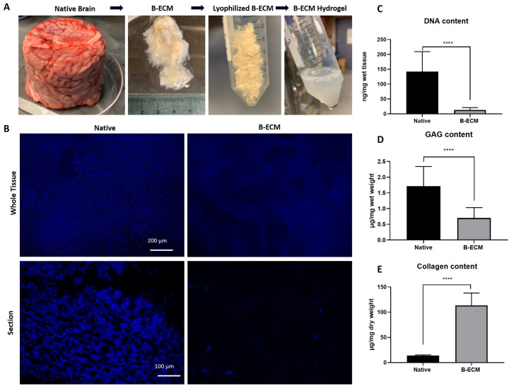
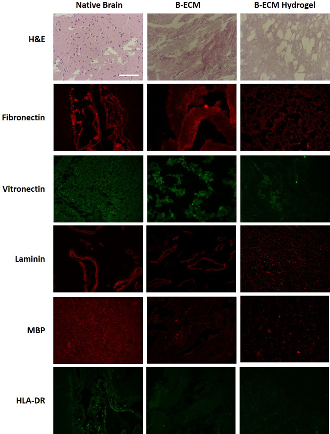
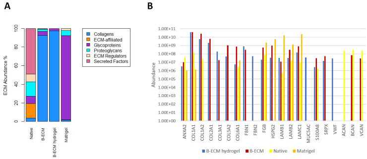
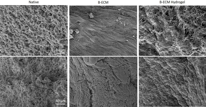
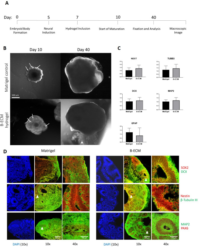
Human brain tissue models such as cerebral organoids are essential tools for developmental and biomedical research. Current methods to generate cerebral organoids often utilize Matrigel as an external scaffold to provide structure and biologically relevant signals. Matrigel however is a nonspecific hydrogel of mouse tumor origin and does not represent the complexity of the brain protein environment. In this study, we investigated the application of a decellularized adult porcine brain extracellular matrix (B-ECM) which could be processed into a hydrogel (B-ECM hydrogel) to be used as a scaffold for human embryonic stem cell (hESC)-derived brain organoids. We decellularized pig brains with a novel detergent- and enzyme-based method and analyzed the biomaterial properties, including protein composition and content, DNA content, mechanical characteristics, surface structure, and antigen presence. Then, we compared the growth of human brain organoid models with the B-ECM hydrogel or Matrigel controls in vitro. We found that the native brain source material was successfully decellularized with little remaining DNA content, while Mass Spectrometry (MS) showed the loss of several brain-specific proteins, while mainly different collagen types remained in the B-ECM. Rheological results revealed stable hydrogel formation, starting from B-ECM hydrogel concentrations of 5 mg/mL. hESCs cultured in B-ECM hydrogels showed gene expression and differentiation outcomes similar to those grown in Matrigel. These results indicate that B-ECM hydrogels can be used as an alternative scaffold for human cerebral organoid formation, and may be further optimized for improved organoid growth by further improving protein retention other than collagen after decellularization.


Conflict of interest statement

The author's have read the journal's policy and the authors of this study have the following competing interests: RS was a paid employee of VERIGRAFT AB at the time of study and HG was a paid employee of Felix Printers at the time of study. This does not alter our adherence to PLOS ONE policies on sharing data and materials. There are no patents, products in development or marketed products associated with this research to declare.

Figures

Fig 1
Fig 1. DNA and ECM content of decellularization pig brain.
(A) Native pig brains were decellularized with detergents, lyophilized, and solubilized in a pepsin-HCl mix. After neutralizing the solution (15 mg/mL) to physiological salt and pH concentration, hydrogel formation at 37°C was observed. (B) DAPI staining of whole tissue samples en face (upper panel) and tissue sections (lower panel). (C) DNA content in native brain and B-ECM samples (n = 9) was measured after extraction of DNA from wet tissue with a commercial kit. (D) GAGs from wet tissue (n = 8) and (E) collagen from dry tissue (n = 15) were quantified by extraction of proteins with commercial kits.
Fig 2
Fig 2. Staining of native, B-ECM and B-ECM hydrogel samples.
Histological and immunostaining was performed on paraffin-embedded samples after rehydration and antigen retrieval (n = 3). For general morphology, the samples were stained with hematoxylin-eosin (H&E). Fibronectin, vitronectin, laminin, myelin basic protein (MBP) and human leucocyte antigen (HLA-DR) were detected with antibodies (immunofluorescence). Scale bar represents 100 μm.
Fig 3
Fig 3. Protein analysis performed by mass spectrometry on samples from native brain, B-ECM, B-ECM hydrogel, and on Matrigel.
(A) Stacked bar chart of the abundance of the matrisome-proteins that were detected in the analyzed samples, categorized in different matrisome categories (Collagens, ECM-affiliated, Glycoproteins, Proteoglycans, ECM Regulators or Secreted Factors) (B) The abundance of ECM proteins found in the B-ECM hydrogel sample compared to Native, B-ECM, and Matrigel samples, sorted by ECM categories (collagens, glycoproteins, and proteoglycans) with their gene identifier. Besides, the abundances of the brain-specific ECM proteins brevican (BCAN), aggrecan (ACAN), and versican (VCAN) in the different samples are shown as well. Y-axis is in the logarithmic scale.
Fig 4
Fig 4. SEM of native, B-ECM and B-ECM hydrogel samples.
Images taken with 4000x (upper panel) and 1000x (lower panel) magnification.
Fig 5
Fig 5. Growth and gene expression of brain organoids embedded in B-ECM hydrogel or Matrigel.
(A) Timeline of experimental steps of human brain organoid maturation embedded in Matrigel or B-ECM hydrogel. (B) Brain organoids (n = 4) on day 10 in Matrigel (top pictures) or B-ECM hydrogel (bottom pictures). On day 40, no significant difference is apparent. White arrows at day 10 point to ventricular-like zones located in the formed neuronal buds White arrowhead at day 40 indicates residual B-ECM hydrogel. Scale bar: 500 μm. (C) Expression of the genes NEST, TUBB3, DCX, MAP2 and GFAP was measured by extraction of RNA from organoids grown in the presence of B-ECM hydrogel (n = 13) or Matrigel (n = 12) in technical triplicates with RT-qPCR. Statistical analysis performed with unpaired t-test. (D) Tissue sections of brain organoids cultured in Matrigel (left panels) or B-ECM hydrogel (right panels) for 40 days were immunostained for neuronal progenitor markers (SOX2, Nestin, PAX6) and mature neuronal markers (DCX, β-Tubulin III, MAP2). Images were taken with a 10x objective (10x, scale bar: 300 μm) and 40x objective (40x, scale bar: 100 μm). DAPI was used as nuclear counterstaining. White arrowheads indicate ventricular-like zones that were chosen for magnification.

Similar articles

Cited by

References

    1. Rothenbücher TSP, Martínez-Serrano A. Human cerebral organoids and neural 3D tissues in basic research, and their application to study neurological diseases Future Neurol. Future Medicine Ltd; London, UK; 2019;14: FNL3 10.2217/fnl-2018-0043 - DOI
    1. Lancaster MA, Renner M, Martin C-A, Wenzel D, Bicknell LS, Hurles ME, et al. Cerebral organoids model human brain development and microcephaly. Nature. 2013;501: 373–379. 10.1038/nature12517 - DOI - PMC - PubMed
    1. Stachowiak EK, Benson CA, Narla ST, Dimitri A, Chuye LEB, Dhiman S, et al. Cerebral organoids reveal early cortical maldevelopment in schizophrenia—computational anatomy and genomics, role of FGFR1 Transl Psychiatry. Nature Publishing Group; 2017;7: 6 10.1038/s41398-017-0054-x - DOI - PMC - PubMed
    1. Qian X, Nguyen HN, Song MM, Hadiono C, Ogden SC, Hammack C, et al. Brain-Region-Specific Organoids Using Mini-bioreactors for Modeling ZIKV Exposure. Cell. 2016;165: 1238–1254. 10.1016/j.cell.2016.04.032 - DOI - PMC - PubMed
    1. Raja WK, Mungenast AE, Lin Y-T, Ko T, Abdurrob F, Seo J, et al. Self-Organizing 3D Human Neural Tissue Derived from Induced Pluripotent Stem Cells Recapitulate Alzheimer’s Disease Phenotypes. Padmanabhan J, editor. PLoS One. 2016;11: e0161969 10.1371/journal.pone.0161969 - DOI - PMC - PubMed

Publication types