. 2021 Jan 28;17(1):e1009255.
doi: 10.1371/journal.ppat.1009255. eCollection 2021 Jan.

Hematopoietic cell-mediated dissemination of murine cytomegalovirus is regulated by NK cells and immune evasion

Affiliations

Hematopoietic cell-mediated dissemination of murine cytomegalovirus is regulated by NK cells and immune evasion

Shunchuan Zhang et al. PLoS Pathog. .

Abstract

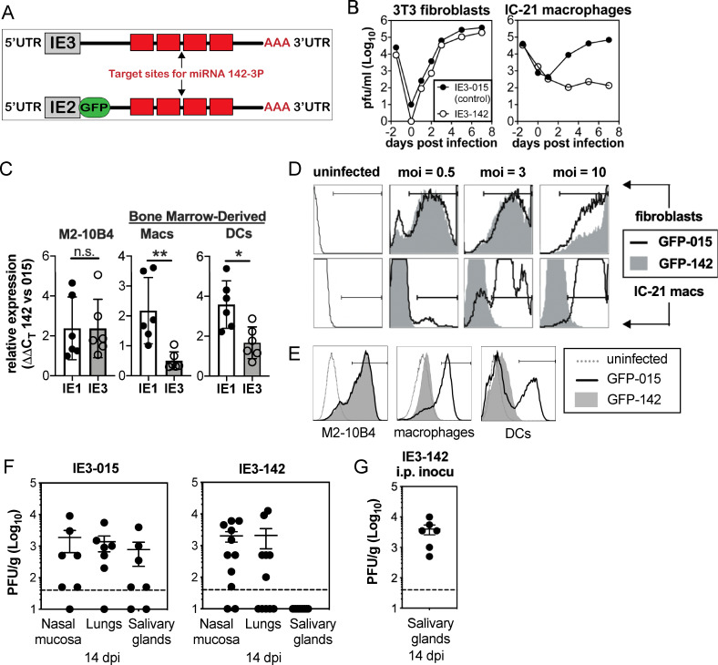
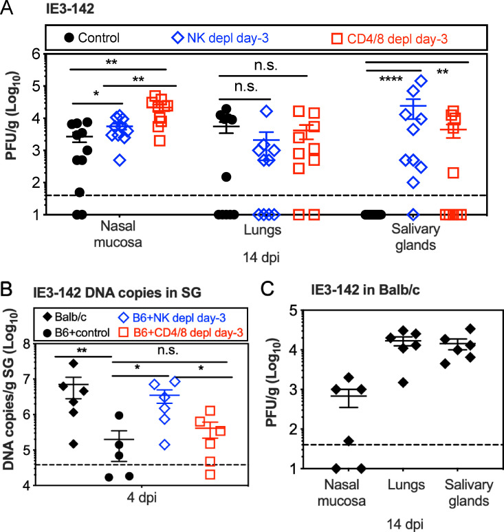
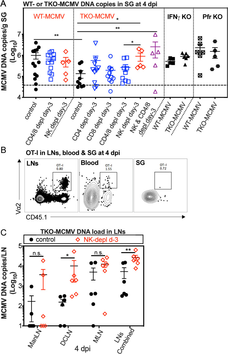
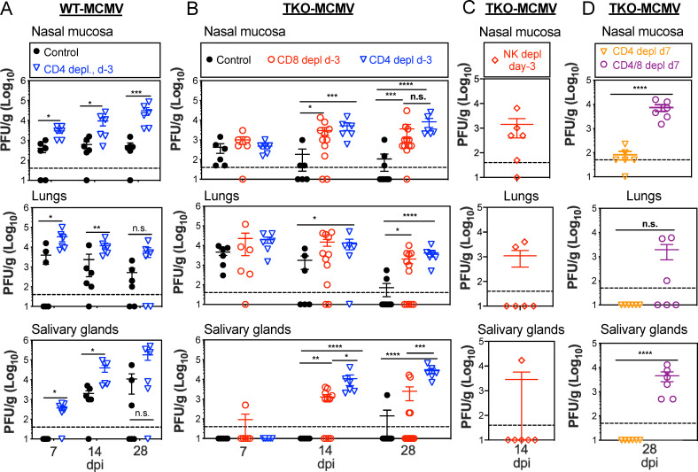
Cytomegalovirus (CMV) causes clinically important diseases in immune compromised and immune immature individuals. Based largely on work in the mouse model of murine (M)CMV, there is a consensus that myeloid cells are important for disseminating CMV from the site of infection. In theory, such dissemination should expose CMV to cell-mediated immunity and thus necessitate evasion of T cells and NK cells. However, this hypothesis remains untested. We constructed a recombinant MCMV encoding target sites for the hematopoietic specific miRNA miR-142-3p in the essential viral gene IE3. This virus disseminated poorly to the salivary gland following intranasal or footpad infections but not following intraperitoneal infection in C57BL/6 mice, demonstrating that dissemination by hematopoietic cells is essential for specific routes of infection. Remarkably, depletion of NK cells or T cells restored dissemination of this virus in C57BL/6 mice after intranasal infection, while dissemination occurred normally in BALB/c mice, which lack strong NK cell control of MCMV. These data show that cell-mediated immunity is responsible for restricting MCMV to hematopoietic cell-mediated dissemination. Infected hematopoietic cells avoided cell-mediated immunity via three immune evasion genes that modulate class I MHC and NKG2D ligands (m04, m06 and m152). MCMV lacking these 3 genes spread poorly to the salivary gland unless NK cells were depleted, but also failed to replicate persistently in either the nasal mucosa or salivary gland unless CD8+ T cells were depleted. Surprisingly, CD8+ T cells primed after intranasal infection required CD4+ T cell help to expand and become functional. Together, our data suggest that MCMV can use both hematopoietic cell-dependent and -independent means of dissemination after intranasal infection and that cell mediated immune responses restrict dissemination to infected hematopoietic cells, which are protected from NK cells during dissemination by viral immune evasion. In contrast, viral replication within mucosal tissues depends on evasion of T cells.


Conflict of interest statement

The authors have declared that no competing interests exist.

Figures

Fig 1
Fig 1. MCMV must use infected hematopoietic cells for dissemination in C57BL/6 mice after i.n. inoculation.
A. Schematic of miR-142-3p targeted viruses (MCMV-IE3-142 and MCMV-GFP-142). Four target sites for miR-142-3p were inserted into the 3’ untranslated region of the essential viral gene IE3 or into a GFP-SIINFEKL fusion construct inserted into the IE2 locus. Control viruses contain shuttle vector sequences without miR-142-3p target sites in the same location. B. Targeting IE3 with miR-142-3p binding sites prevents viral replication in miR-142-3p-expressing macrophages. Multi-step growth curves of the MCMV-IE3-015 control virus and the MCMV-IE3-142 virus in 3T3 fibroblasts and IC-21 macrophages. C. The miR-142-3p targets IE3 gene and inhibits its gene expression. IE1 and IE3 gene expression were evaluated by RT-PCR after infection of M2-10B4 cells and bone marrow-derived macrophages and dendritic cells with either MCMV-IE3-015 or MCMV-IE3-142 viruses. Gene expression was normalized to β-actin in each sample and compared between MCMV-IE3-142 and MCMV-IE3-015 infected samples using the ΔΔCT method. The bar graph represents the mean value. Each symbol represents an individual cell sample, and error bars represent the standard deviation. Data are from one representative experiment of three independent experiments. D-E. Targeting IE3 with miR-142-3p binding sites inhibits gene expression. Regulation of gene expression by miR-142-3p is visualized by GFP expression after infection of 3T3 fibroblasts and IC-21 macrophages (D) or M2-10B4 cells and bone marrow-derived macrophages and dendritic cells (E) with either MCMV-GFP-015 or MCMV-GFP-142 viruses. F-G. Productive infection of hematopoietic cells is necessary for viral dissemination after i.n. inoculation, but not after i.p. inoculation. Virus titers in the nasal mucosa, lungs and salivary glands at 14 days after i.n. inoculation (F) or i.p. inoculation (G), with 106 PFU MCMV-IE3-142, or control virus MCMV-IE3-015. Each symbol represents an individual animal. The solid line shows the mean titer, and error bars represent the SEM. Dashed lines show the detection limit (50 PFU/g). Data are combined from two independent experiments.
Fig 2
Fig 2. NK cell and T cell work together to modulate virus dissemination.
A. Depletion of NK cells or T cells from C57BL/6 mice enables MCMV-IE3-142 dissemination from the nasal mucosa to the salivary gland. C57BL/6 mice were depleted of NK cells or CD4+ T cells and CD8+ T cells before i.n. infection with MCMV-IE3-142. Shown are the viral titers in the nasal mucosa, lungs and salivary gland 14 days after infection. Each symbol represents an individual animal. The solid line shows the mean value, and error bars represent the SEM. Dashed lines show the detection limit (50 PFU/g). Data are combined from at least two independent experiments. B. Early viral dissemination is affected by NK cell responses. C57BL/6 mice with or without depletion of either NK cells or CD4+ and CD8+ T cells were i.n. inoculated with MCMV-IE3-142. Shown are viral DNA copies in the salivary gland at 4 dpi. Data are displayed as in A and are combined from two independent experiments. C. MCMV does not require infection of hematopoietic cells to reach the salivary glands after i.n. infection of BALB/c mice. Shown are virus titers in the nasal mucosa, lungs and salivary glands of BALB/c mice at 14 days after i.n. inoculation of MCMV-IE3-142. Data are displayed as in A and are combined from two independent experiments.
Fig 3
Fig 3. Early dissemination of TKO-MCMV is restored by NK cell depletion.
A. NK cells prevent early dissemination of TKO-MCMV to the salivary gland after i.n. infection of C57BL/6 mice in a manner dependent on both IFN-γ and perforin. C57BL/6 mice depleted of the indicated cells before i.n. infection or mice lacking IFN-γ or perforin were intranasally inoculated with either WT-MCMV or TKO-MCMV. Shown are viral DNA copies in the salivary gland 4 days after infection. Each symbol represents an individual animal. The solid line shows the mean value, and error bars represent the SEM. Dashed lines show the detection limit. Data are combined from at least two independent experiments for each condition. B. MCMV-specific T cells are not present in the salivary gland by day 4 after i.n. infection. Representative FACS plots show OT-I cells in the blood, draining LNs (ManLNs, DCLNs and MLNs) and salivary gland 4 days after i.n. infection with MCMV-Ova. Data show cells in one representative mouse from one experiment. C. NK cell depletion increases the TKO-MCMV DNA loads in draining LNs (ManLNs, DCLNs and MLNs). NK cells were depleted or not from C57BL/6 mice beginning 3 days prior to infection. Shown are viral DNA copies in the indicated lymph nodes individually or combined at 4 days post infection. Data are displayed as in A and are combined from 2 independent experiments.
Fig 4
Fig 4. Evasion of MHC-I antigen-presentation and CD8+ T cells is critical for viral persistence at site of entry and replication in the salivary gland.
A. WT-MCMV persists in the nasal mucosa and spreads to the salivary gland within 14 days after i.n. infection. Virus titers in the nasal mucosa, lungs and salivary glands of C57BL/6 mice with or without CD4+ T cells depletion before infection at 7, 14 and 28 days after i.n. inoculation of WT-MCMV. Each symbol represents an individual animal. The solid line shows the mean titer, and error bars represent the SEM. Dashed lines show the detection limit (50 PFU/g). Data are combined from two independent experiments. B. Lack of MHC-I evasion genes prevents viral persistence in the nasal mucosa and spread to the salivary gland. Viral titers in the nasal mucosa, lungs and salivary gland 7, 14 and 28 days after i.n. inoculation of C57BL/6 mice infected with TKO-MCMV. Infected mice were treated with an isotype control antibody or depleted of CD4+ or CD8+ T cells before infection. Data are displayed as in A and are combined from at least two independent experiments. C. NK cell depletion before infection does not rescue TKO-MCMV replication in the salivary glands after i.n. infection of C57BL/6 mice. Shown are virus titers in the nasal mucosa, lungs and salivary glands at 14 days post infection. Data are displayed as in A and are combined from two independent experiments. D. CD8+ T cells can control TKO-MCMV if they are primed in the presence of CD4+ T cell help. Virus titers in the indicated organs at day 28 post infection are shown. C57BL/6 mice were depleted of either CD4+ T cells or both CD4+ and CD8+ T cells, beginning at day 7 after i.n. infection of TKO-MCMV. Data are displayed as in A and are combined from two independent experiments.
Fig 5
Fig 5. CD4+ T cell help is needed for the proliferation and function of MCMV-specific CD8+ T cells.
A. CD8+ T cells are reduced in frequency after i.n. infection in the absence of CD4+ T cell help. Shown is the frequency of viral tetramer-specific CD8+ T cells in the blood of recipients at the indicated time points with or without CD4+ T cell depletion before infection. Data show the average frequency of T cells at day 7 (n = 9–12), day 14 (n = 6–9) and day 28 (n = 3–6) after i.n. infection and are derived from one representative experiment of at least 3 independent experiments. B. CD8+ T cell function is impaired in the absence of CD4+ T cell help after i.n. infection, but improved by delaying CD4+ T cell depletion until day 7. Each symbol represents an individual animal. The solid line shows the mean value, and error bars represent the SEM. Data are combined from two independent experiments. C. The frequency and number of OT-Is are impaired in the absence of CD4+ T cell help after i.n. infection with MCMV-Ova. CD4+ T cells were depleted or not from C57BL/6 mice. One day before i.n. infection with MCMV-Ova, mice received 5000 OT-I T cells. Shown are the frequency (left) and absolute number (right) of OT-I cells in blood over time after infection. Data show the average values from 6–8 animals per group, and error bars represent the SEM. CountBright absolute counting beads (ThermoFisher Scientific) were included to determine the number of cells in the blood. D. The function of OT-I T cells in the spleen is impaired on a per-cell basis in the absence of CD4+ T cell help. OT-I T cells in the spleens of adoptive recipients (as described in C.) were analysed for production of IFN-γ, TNF-α and degranulation (exposure of CD107a) after stimulation with SIINFEKL peptide 28 days after infection. Each symbol represents an individual animal. The solid line shows the mean value, and error bars represent the SEM.

Similar articles

Cited by

References

    1. Mussi-Pinhata M, Yamamoto A, Moura Brito R, de Lima Isaac M, de Carvalho e Oliveira P, Boppana S, et al. Birth prevalence and natural history of congenital cytomegalovirus infection in a highly seroimmune population. Clin Infect Dis. 2009;49(4):522–8. 10.1086/600882 . - DOI - PMC - PubMed
    1. Manicklal S, Emery V, Lazzarotto T, Boppana S, Gupta R. The "silent" global burden of congenital cytomegalovirus. Clin Microbiol Rev. 2013;26(1):86–102. 10.1128/CMR.00062-12 . - DOI - PMC - PubMed
    1. Kenneson A, Cannon M. Review and meta-analysis of the epidemiology of congenital cytomegalovirus (CMV) infection. Rev Med Virol. 2007;17(4):253–76. 10.1002/rmv.535 . - DOI - PubMed
    1. Bhide A, Papageorghiou AT. Managing primary CMV infection in pregnancy. BJOG. 2008;115(7):805–7. 10.1111/j.1471-0528.2008.01728.x . - DOI - PubMed
    1. Farrell HE, Lawler C, Tan CS, MacDonald K, Bruce K, Mach M, et al. Murine Cytomegalovirus Exploits Olfaction To Enter New Hosts. mBio. 2016;7(2):e00251–16. 10.1128/mBio.00251-16 - DOI - PMC - PubMed

Publication types

MeSH terms