. 2021 Jul 1;23(7):1087-1099.
doi: 10.1093/neuonc/noab012.

Genome-wide methylation profiling of glioblastoma cell-derived extracellular vesicle DNA allows tumor classification

Affiliations

Genome-wide methylation profiling of glioblastoma cell-derived extracellular vesicle DNA allows tumor classification

Cecile L Maire et al. Neuro Oncol. .

Abstract

Background: Genome-wide DNA methylation profiling has recently been developed into a tool that allows tumor classification in central nervous system tumors. Extracellular vesicles (EVs) are released by tumor cells and contain high molecular weight DNA, rendering EVs a potential biomarker source to identify tumor subgroups, stratify patients and monitor therapy by liquid biopsy. We investigated whether the DNA in glioblastoma cell-derived EVs reflects genome-wide tumor methylation and mutational profiles and allows noninvasive tumor subtype classification.

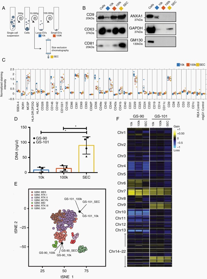
Methods: DNA was isolated from EVs secreted by glioblastoma cells as well as from matching cultured cells and tumors. EV-DNA was localized and quantified by direct stochastic optical reconstruction microscopy. Methylation and copy number profiling was performed using 850k arrays. Mutations were identified by targeted gene panel sequencing. Proteins were differentially quantified by mass spectrometric proteomics.

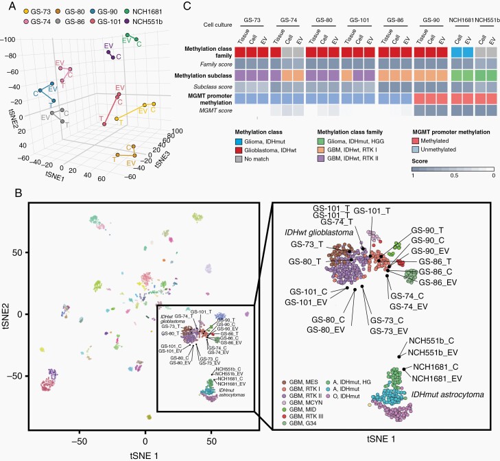
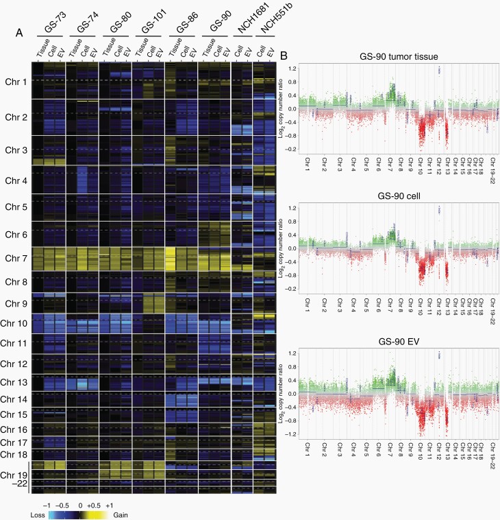
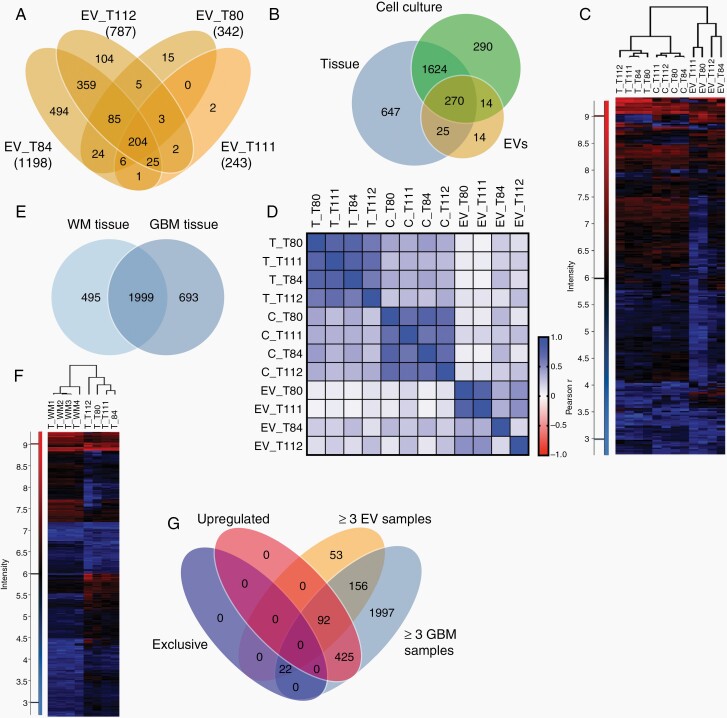
Results: Genome-wide methylation profiling of glioblastoma-derived EVs correctly identified the methylation class of the parental cells and original tumors, including the MGMT promoter methylation status. Tumor-specific mutations and copy number variations (CNV) were detected in EV-DNA with high accuracy. Different EV isolation techniques did not affect the methylation profiling and CNV results. DNA was present inside EVs and on the EV surface. Proteome analysis did not allow specific tumor identification or classification but identified tumor-associated proteins that could potentially be useful for enriching tumor-derived circulating EVs from biofluids.

Conclusions: This study provides proof of principle that EV-DNA reflects the genome-wide methylation, CNV, and mutational status of glioblastoma cells and enables their molecular classification.

Keywords: exosome; glioma; methylome; mutation; proteomics.


Figures

Fig. 1
Fig. 1
Extracellular vesicle (EV) isolation and characterization. A, Glioma cells release small EVs from multivesicular bodies (MVB), coordinated by the endosomal sorting complex required for transport (ESCRT), while larger EVs are generated by membrane budding. Glioma tissue was cultured and EVs secreted by tumor cells were analyzed. B, Electron microscopy demonstrates the cup-shaped morphology of EVs (arrows). C, Detection of CD9, CD63, and CD81 by imaging flow cytometry. D, Direct stochastic optical reconstruction microscopy of EVs identified by CD63 or CD81 and stained with nucleic acid dye. DNase treatment removed extra-vesicular DNA, and permeabilization accessed intra-vesicular DNA. E, Quantification of the number of DNA fragment localizations per EV, based on co-localization with CD63 and/or CD81. Box plots with 10-90 percentile (whiskers), median (line) and 25-75 percentile (box); ****P < .0001, Kruskal-Wallis analysis.
Fig. 2
Fig. 2
DNA methylation profiling of EV-DNA. A, t-SNE analysis of genome-wide methylation profiles. B, Comparison of samples to the CNS tumor reference cohort (left). All samples from IDHwt tumors EVs, cells (C), and tumors (T), cluster in close spatial proximity to RTKI and RTKII reference clusters, while cells and EVs from IDHmut tumors cluster with IDH-mutant high-grade astrocytomas (right, magnified glioma clusters). C, Summarized results of methylation profiling reports. Abbreviations: EV, extracellular vesicles; IDHwt, isocitrate dehydrogenase wild-type; t-SNE, t-distributed stochastic neighbor embedding dimensionality reduction.
Fig. 3
Fig. 3
CNV analysis of EV-DNA. A, Heatmap representation of genome-wide copy number gains and losses inferred from the DNA methylation analysis. B, Example of CNV profiles for tumor T90 with corresponding cells and EVs. Abbreviations: CNV, copy number variations; EV, extracellular vesicles.
Fig. 4
Fig. 4
Mutation profiles of EV-DNA. The variant allele frequency (VAF) was determined by NGS. *TERT promoter mutations were analyzed by ddPCR since NGS reads are usually low in this region. Abbreviations: ddPCR, digital droplet PCR; NGS, next-generation sequencing.
Fig. 5
Fig. 5
Proteomic profiles of glioma extracellular vesicles (EVs). A, Numbers of proteins detected in EVs from cultured cells of four glioblastomas by differential quantitative proteomics. B, Proteins detected in at least three of four samples each of EVs, cells, and original tumors. C, Unsupervised clustering based on 270 proteins present in all three sample types. D, Pearson correlation analysis of the proteomes of EVs, cells, (C) and tumor tissue (T). E, Overlap between proteins presents in at least three of four white matter (WM) or glioblastoma (GBM) samples. F, Unsupervised hierarchical clustering based on proteins (2172) detected in GBMs and WM. G, Proteins either exclusively detected in GBM tissue, upregulated in GBMs vs WM (≥2-fold), present in ≥3EV samples and in ≥3 GBMs.
Fig. 6
Fig. 6
EV-DNA methylation profiling is independent of isolation techniques. A, EVs were isolated by centrifugation at 10,000 × g (10k), ultracentrifugation (100k), or size exclusion chromatography (SEC). B, Immunoblot analysis of GS-90 cells and EVs. C, Characterization of EV surface proteins by multiplex bead-based flow cytometry analysis (GS-57, GS-73, GS-74, GS-90, GS-101). D, DNA amount in EVs measured by qBit (GS-90, n = 3, GS-101, n = 1), ANOVA analysis. Values are means ± SD, *P < .05. E, Methylation profiling of EV-DNA analyzed in comparison to the CNS tumor reference cohort. F, Heatmap representation of genome-wide CNV inferred from the DNA methylation analysis. Abbreviations: CNV, copy number variations; EV, extracellular vesicles.

References

    1. Ricklefs FL, Maire CL, Reimer R, et al. . Imaging flow cytometry facilitates multiparametric characterization of extracellular vesicles in malignant brain tumours. J Extracell Vesicles. 2019;8(1):1588555. - PMC - PubMed
    1. Osti D, Del Bene M, Rappa G, et al. . Clinical significance of extracellular vesicles in plasma from glioblastoma patients. Clin Cancer Res. 2019;25(1):266–276. - PubMed
    1. Jeppesen DK, Fenix AM, Franklin JL, et al. . Reassessment of exosome composition. Cell. 2019;177(2):428–445.e18. - PMC - PubMed
    1. Akers JC, Ramakrishnan V, Kim R, et al. . miRNA contents of cerebrospinal fluid extracellular vesicles in glioblastoma patients. J Neurooncol. 2015;123(2):205–216. - PMC - PubMed
    1. Ebrahimkhani S, Vafaee F, Hallal S, et al. . Deep sequencing of circulating exosomal microRNA allows non-invasive glioblastoma diagnosis. NPJ Precis Oncol. 2018;2:28. - PMC - PubMed

Publication types