. 2021 May 6;29(5):499-506.e3.
doi: 10.1016/j.str.2021.01.004. Epub 2021 Jan 27.

Predictable cholesterol binding sites in GPCRs lack consensus motifs

Affiliations

Predictable cholesterol binding sites in GPCRs lack consensus motifs

Geoffrey J Taghon et al. Structure. .

Abstract

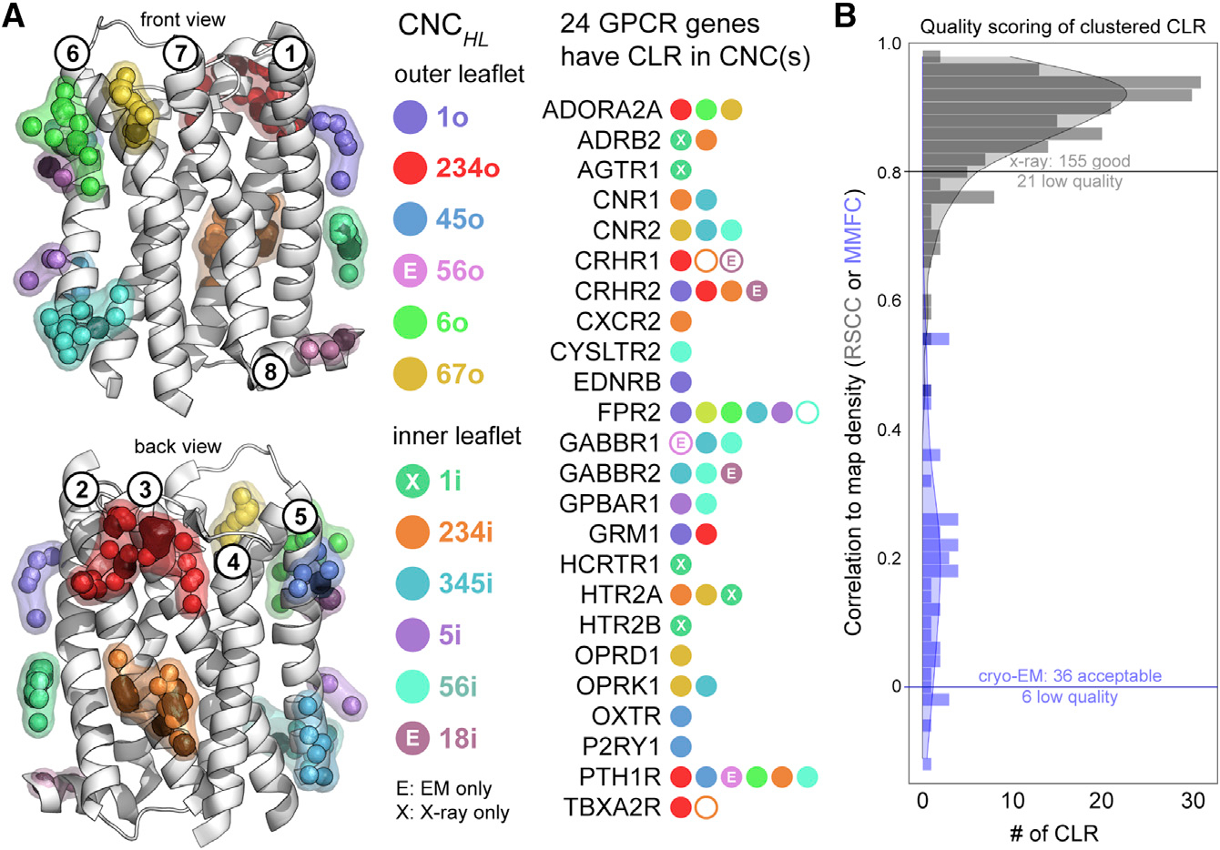
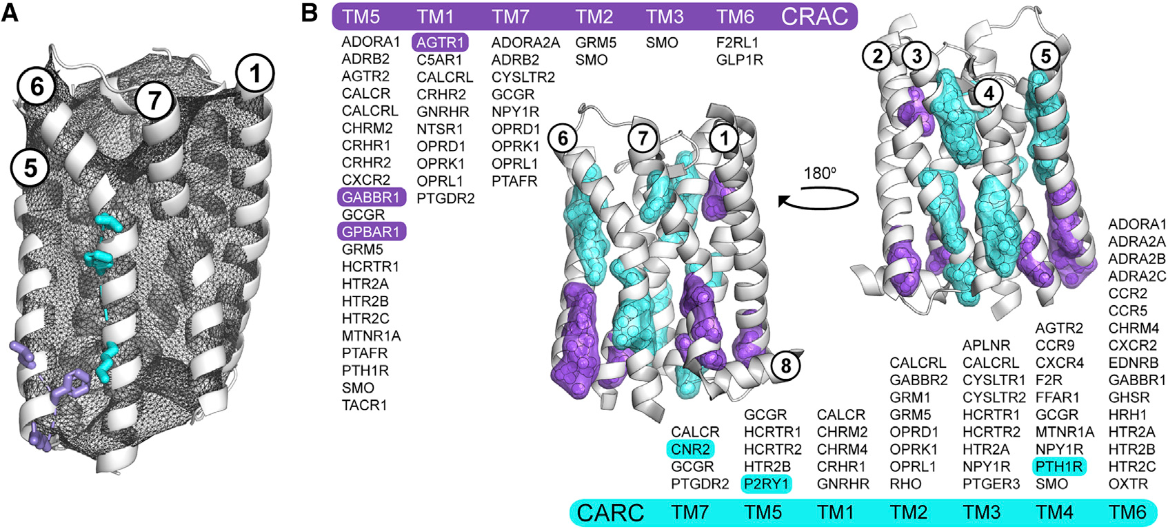
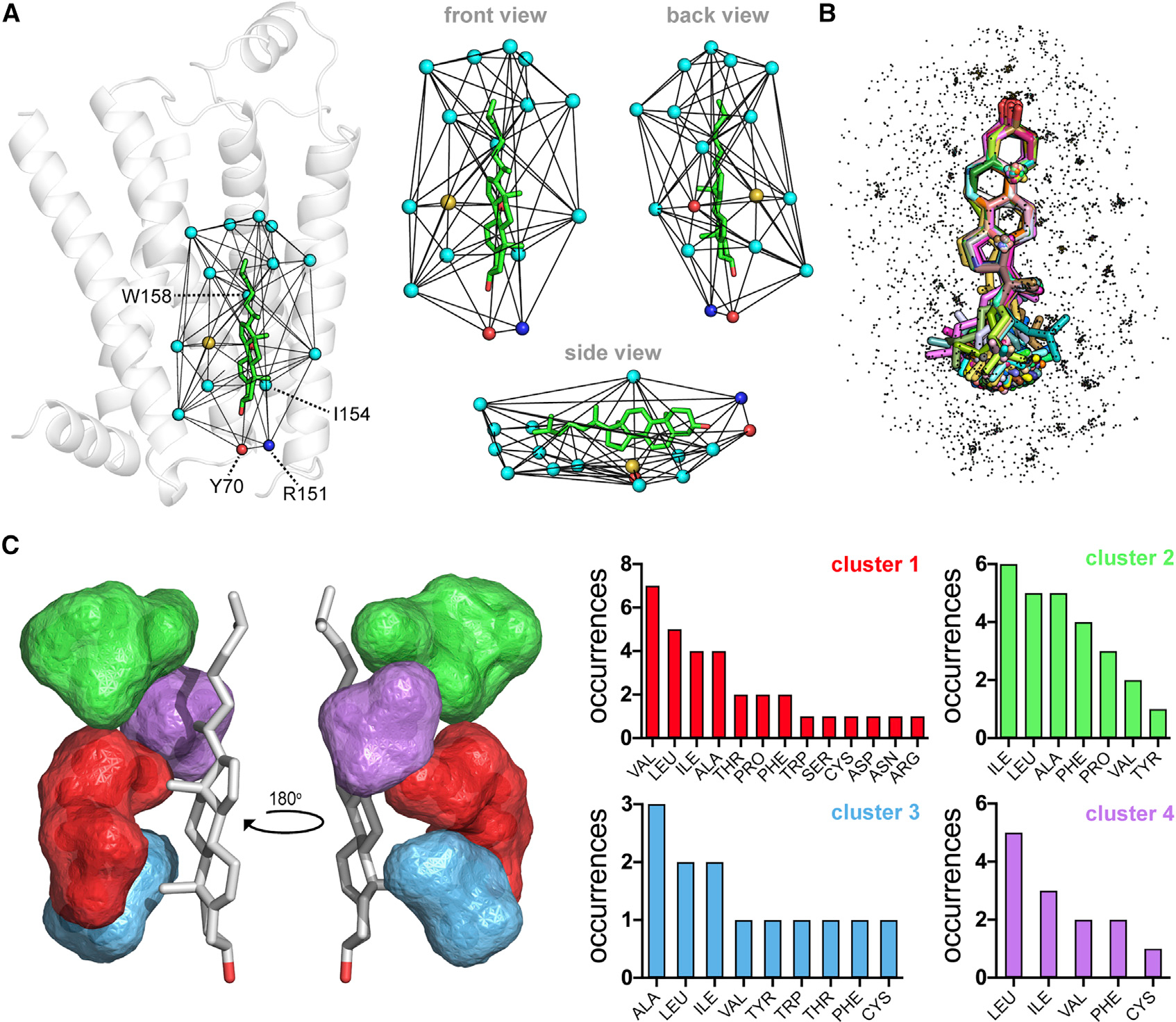
A rich diversity of transmembrane G protein-coupled receptors (GPCRs) are used by eukaryotes to sense physical and chemical signals. In humans alone, 800 GPCRs comprise the largest and most therapeutically targeted receptor class. Recent advances in GPCR structural biology have produced hundreds of GPCR structures solved by X-ray diffraction and increasingly, cryo-electron microscopy (cryo-EM). Many of these structures are stabilized by site-specific cholesterol binding, but it is unclear whether these interactions are a product of recurring cholesterol-binding motifs and if observed patterns of cholesterol binding differ by experimental technique. Here, we comprehensively analyze the location and composition of cholesterol binding sites in the current set of 473 human GPCR structural chains. Our findings establish that cholesterol binds similarly in cryo-EM and X-ray structures and show that 92% of cholesterol molecules on GPCR surfaces reside in predictable locations that lack discernable cholesterol-binding motifs.

Keywords: G protein-coupled receptors (GPCRs); X-ray diffraction; cholesterol; cholesterol consensus motif (CCM); cholesterol recognition/interaction amino acid consensus (CRAC) motif; cryo-electron microscopy (cryo-EM); lipids; structural informatics.


Conflict of interest statement

Declaration of interests The authors declare that they have no conflicts of interest with the contents of this article.

Figures

Figure 1.
Figure 1.. Structural informatics of lipids bound to human GPCR surfaces
(A–C) Breakdown of the current set of 473 human GPCR structural chains by method (A) and their most prevalent bound lipids (B). Overview of the pHinder algorithm for calculating CNCs (C). First, 240 surface-bound CLRs were collected from 473 GPCR structures aligned to the reference receptor bovine rhodopsin (RHO, PDB code 1F88, chain A) (C, left). The set of CLRs were then reduced to geometric centroids, triangulated, and clustered (C, right). The full set of 240 CLR centroids is colored purple. The subset of CLR centroids clustered in CNCs (221 of 240 nodes) are colored red, and the subset of CLR centroids that did not cluster to CNCs (19 of 240 nodes) are colored green. Additional details regarding the set of 473 human GPCR structures, CLR collection procedure, and CAN algorithm are available in STAR methods and Table S1.
Figure 2.
Figure 2.. Cholesterol Network Clusters in human GPCRs
(A and B) Two hundred and twenty-one of 240 (92%) CLR molecules in human GPCR structures map to one of 12 spatially distinct CNCs. Of the 81 unique GPCR genes that contribute the set of 473 GPCR structures, 24 have CLR(s) located in CNCs described using the nomenclature CNCHL, where the H subscript corresponds to the transmembrane helix or helices contacted by the CNC, and the L subscript indicates whether the CNC is located in the inner or outer leaflet of the membrane bilayer. CNCs labeled with an X or E correspond to CLR clusters observed exclusively in X-ray or cryo-EM structures, respectively (A). RSCC (gray) and MMFC (blue) scoring for the set of 221 CLR that mapped to CNCs (B). Low-quality CLRs that mapped to CNCs are indicated as black centroids (A, left) and empty circles in the list of 24 GPCR genes (A, right).
Figure 3.
Figure 3.. CRAC/CARC motifs are not predictive of cholesterol binding in human GPCRs
(A and B) Overview of the procedure for validating the location and directionality of CRAC/CARC motifs in GPCRs using the adenosine A1 receptor (ADORA1, PDB code 5N2S, chain A) and two of its putative CRAC (L201, F204, R208; purple) and CARC (K234, F241, L245; cyan) motifs (A). Valid CRAC/CARC motifs met two geometric criteria: the three side chains comprising the motif are located outside the pHinder surface (shown in black mesh) and oriented in the same general direction. Triangulation and clustering of the 164 and 186 putative CRAC (purple) and CARC (cyan) motifs in 473 GPCR structural chains (B). Each motif was first converted to a geometric centroid prior to triangulating and clustering with a minimum cluster size of five. The subset of GPCRs with validated CRAC/CARC motifs are listed by transmembrane helix (TM). Only six of these motifs (highlighted) had CLR observed nearby in a structure. CRAC clusters containing contributions from TM3 (SMO) and TM6 (F2RL1, GLP1R), and CARC clusters containing contributions from TM1 (CACLR, CHRM2, CHRM4, GNRHR) are not shown because they did not meet the minimum cluster size.
Figure 4.
Figure 4.. Clustered CLR microenvironments in GPCRs lack distinct CLR-binding motifs
(A–C) Overview of the triangulation and extraction procedure for collecting CLR microenvironments (A). Triangulation of the CLR (CLR 402, PDB code 3D4S, chain A) used to classify the CCM, indicated by the residue labels Y70, R151, I154, and W158, in an early structure of the prototypical β2-adrenergic receptor. Spheres represent the TSC atom of each residue in the first CLR triangulation shell and are colored by atom type (cyan, carbon; yellow, sulfur; blue, nitrogen; red, oxygen). Composite overlay of 217 aligned CLR (sticks) and their TSC atom microenvironments (dots) (B). Triangulation and clustering of the combined CLR microenvironment data shown in (B), but with redundant structural information removed (C). Cluster locations and composition bar charts are matched by color. The low-quality CLR indicated in Figure 2B were removed from this analysis, reducing the number of CLRs in the set from 240 to 217 (B and C).

Similar articles

Cited by

References

    1. Caffrey M (2015). A comprehensive review of the lipid cubic phase or in meso method for crystallizing membrane and soluble proteins and complexes. Acta Crystallogr. F Struct. Biol. Commun. 71, 3–18. - PMC - PubMed
    1. Cherezov V, Rosenbaum DM, Hanson MA, Rasmussen SG, Thian FS, Kobilka TS, Choi HJ, Kuhn P, Weis WI, Kobilka BK, et al. (2007). High-resolution crystal structure of an engineered human beta2-adrenergic G protein-coupled receptor. Science 318, 1258–1265. - PMC - PubMed
    1. Di Scala C, Baier CJ, Evans LS, Williamson PTF, Fantini J, and Barrantes FJ (2017). Relevance of CARC and CRAC cholesterol-recognition motifs in the nicotinic acetylcholine receptor and other membrane-bound receptors. Curr. Top. Membr. 80, 3–23. - PubMed
    1. Duncan AL, Song W, and Sansom MSP (2020). Lipid-dependent regulation of ion channels and G protein-coupled receptors: insights from structures and simulations. Annu. Rev. Pharmacol. Toxicol. 60, 31–50. - PubMed
    1. Dunn HA, Orlandi C, and Martemyanov KA (2019). Beyond the ligand: extracellular and transcellular G protein-coupled receptor complexes in physiology and pharmacology. Pharmacol. Rev. 71, 503–519. - PMC - PubMed

Publication types