. 2021 Feb 4;108(2):357-367.
doi: 10.1016/j.ajhg.2021.01.008. Epub 2021 Jan 27.

De novo TRIM8 variants impair its protein localization to nuclear bodies and cause developmental delay, epilepsy, and focal segmental glomerulosclerosis

Patricia L Weng  1 Amar J Majmundar  2 Kamal Khan  3 Tze Y Lim  4 Shirlee Shril  2 Gina Jin  4 John Musgrove  5 Minxian Wang  6 Dina F Ahram  4 Vimla S Aggarwal  7 Louise E Bier  8 Erin L Heinzen  8 Ana C Onuchic-Whitford  9 Nina Mann  2 Florian Buerger  2 Ronen Schneider  2 Konstantin Deutsch  2 Thomas M Kitzler  2 Verena Klämbt  2 Amy Kolb  2 Youying Mao  2 Christelle Moufawad El Achkar  10 Adele Mitrotti  4 Jeremiah Martino  4 Bodo B Beck  11 Janine Altmüller  12 Marcus R Benz  13 Shoji Yano  14 Mohamad A Mikati  15 Talha Gunduz  15 Heidi Cope  16 Vandana Shashi  16 Undiagnosed Diseases NetworkHoward Trachtman  17 Monica Bodria  18 Gianluca Caridi  18 Isabella Pisani  19 Enrico Fiaccadori  19 Asmaa S AbuMaziad  20 Julian A Martinez-Agosto  21 Ora Yadin  1 Jonathan Zuckerman  22 Arang Kim  23 UCLA Clinical Genomics CenterUlrike John-Kroegel  24 Amanda V Tyndall  25 Jillian S Parboosingh  25 A Micheil Innes  25 Agnieszka Bierzynska  26 Ania B Koziell  27 Mordi Muorah  28 Moin A Saleem  26 Julia Hoefele  29 Korbinian M Riedhammer  30 Ali G Gharavi  4 Vaidehi Jobanputra  31 Emma Pierce-Hoffman  32 Eleanor G Seaby  32 Anne O'Donnell-Luria  33 Heidi L Rehm  32 Shrikant Mane  34 Vivette D D'Agati  35 Martin R Pollak  36 Gian Marco Ghiggeri  18 Richard P Lifton  37 David B Goldstein  8 Erica E Davis  38 Friedhelm Hildebrandt  39 Simone Sanna-Cherchi  40
Affiliations

De novo TRIM8 variants impair its protein localization to nuclear bodies and cause developmental delay, epilepsy, and focal segmental glomerulosclerosis

Patricia L Weng et al. Am J Hum Genet. .

Abstract

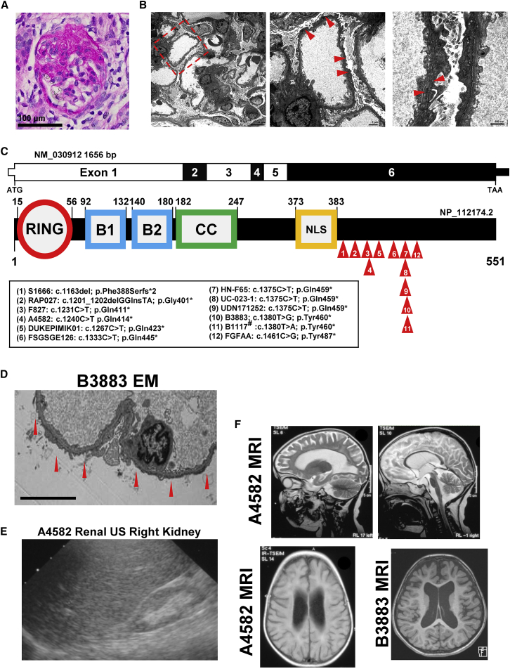
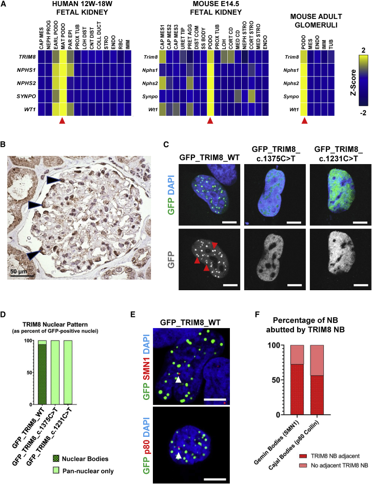
Focal segmental glomerulosclerosis (FSGS) is the main pathology underlying steroid-resistant nephrotic syndrome (SRNS) and a leading cause of chronic kidney disease. Monogenic forms of pediatric SRNS are predominantly caused by recessive mutations, while the contribution of de novo variants (DNVs) to this trait is poorly understood. Using exome sequencing (ES) in a proband with FSGS/SRNS, developmental delay, and epilepsy, we discovered a nonsense DNV in TRIM8, which encodes the E3 ubiquitin ligase tripartite motif containing 8. To establish whether TRIM8 variants represent a cause of FSGS, we aggregated exome/genome-sequencing data for 2,501 pediatric FSGS/SRNS-affected individuals and 48,556 control subjects, detecting eight heterozygous TRIM8 truncating variants in affected subjects but none in control subjects (p = 3.28 × 10⁻¹¹). In all six cases with available parental DNA, we demonstrated de novo inheritance (p = 2.21 × 10⁻¹⁵). Reverse phenotyping revealed neurodevelopmental disease in all eight families. We next analyzed ES from 9,067 individuals with epilepsy, yielding three additional families with truncating TRIM8 variants. Clinical review revealed FSGS in all. All TRIM8 variants cause protein truncation clustering within the last exon between residues 390 and 487 of the 551 amino acid protein, indicating a correlation between this syndrome and loss of the TRIM8 C-terminal region. Wild-type TRIM8 overexpressed in immortalized human podocytes and neuronal cells localized to nuclear bodies, while constructs harboring patient-specific variants mislocalized diffusely to the nucleoplasm. Co-localization studies demonstrated that Gemini and Cajal bodies frequently abut a TRIM8 nuclear body. Truncating TRIM8 DNVs cause a neuro-renal syndrome via aberrant TRIM8 localization, implicating nuclear bodies in FSGS and developmental brain disease.

Keywords: FSGS; SRNS; TRIM8; epilepsy; genomics; monogenic; nuclear body.


Conflict of interest statement

F.H. is a co-founder of Goldfinch Biopharma Inc. The other authors declare that they have no competing financial interests. No part of this manuscript has been previously published.

Figures

Figure 1
Figure 1
Truncating TRIM8 mutations identified in 12 families with nephrotic syndrome and neurologic disease (A) Periodic acid Schiff (PAS) staining of paraffin sections of kidney cortex from a renal biopsy of the index case subject UC-023-1 was performed. A representative glomerulus shows a lesion of focal segmental glomerulosclerosis with segmental obliteration of capillary lumina by extracellular matrix accompanied by loss of overlying podocytes and broad adhesions to Bowman’s capsule (PAS, scale bar: 100 μm). (B) Ultrastructural analysis of kidney tissue from a renal biopsy of the index case UC-023-1 was performed by transmission electron microscopy. A low-power view (left, 2,500×) of the glomerular capillaries shows patent lumina and unremarkable mesangium. There is nearly complete (>90%) effacement of the podocyte foot processes with microvillous transformation of the podocyte cytoplasm. A higher-power view (middle, 6,300×) shows a glomerular capillary with thinning and irregularities of the glomerular basement membrane as well as marked foot process effacement with microvillous change. There are no immune-type electron dense deposits. In the highest-power view (right, 16,000×), two adjacent glomerular capillaries show thinning (124–131 nm) of their glomerular basement membranes accompanied by textural irregularities with focal mild lamellation (arrowhead). Scale bars: 4 μm in left, 1 μm in middle, 600 nm in right. (C) Coding exon (upper bar) and protein domain (lower bar) structures of TRIM8 are shown with arrows indicating position of heterozygous mutations identified in nine subjects. B1, B-box domain 1; B2, B-box domain 2; CC, coiled-coil domain; NLS, nuclear localization signal; RING, ring finger domain. #This patient was independently recruited and published, while the current manuscript was in preparation. (D) Renal biopsy was performed in subject B3883 at age 8 years. Ultrastructural findings by electron microscopy (EM) at 3,000× include diffuse podocyte foot process effacement (red arrows) and thin glomerular basement membranes. Scale bar: 6 μm. (E) Renal ultrasound (RUS) is shown for subject A4582 at 2.75 years, demonstrating a hyperechogenic and atrophied (4.68 cm) right kidney after progression to ESRD. (F) Brain magnetic resonance imaging is shown for subject A4582 at 2.75 years and B3883 at 6 years of life. T2 transverse images of A4582 show deep sulci, atrophied gyri, and thin corpus callosum (top left) and atrophy of corpus callosum, pons and midbrain, while cerebellum preserved (top right). T2 flare axial image of A4582 (bottom left) shows atrophied white matter and hyperintense periventricular signal intensity. T1 axial image of B3883 shows increased ventricle size and sulcal spaces.
Figure 2
Figure 2
TRIM8 is expressed in podocytes of mammalian kidneys and localizes to nuclear bodies (A) TRIM8 mRNA (z-score) was predominantly expressed by podocytes (red arrows) from single-cell mRNA sequencing data.,, In human fetal kidneys from 12 to 19 weeks gestation (left), TRIM8 expression was highest in the mature podocyte cluster, marked by expression of NPHS1, NPHS2, SYNPO, and WT1, relative to other developmental cell-type and nephron segment clusters. Trim8 expression in E14.5 mouse kidneys (middle) was, similarly, highest in the podocyte cluster (Nphs1, Nphs2, Synpo, Wt1) with lower expression in one cap mesenchyme cluster and two tubular segment clusters. In adult mouse glomeruli (right), Trim8 mRNA was predominantly expressed in podocytes relative to other clusters. CAP MES, cap mesenchyme; CNT DIST, distal connecting tubule; COLL DUCT, collecting duct; CORT CD, cortical collecting duct; CORT STRO, cortical stroma; DIST COM, distal comma shaped body; EARL PODO, early podocyte; ENDO, endothelium; IMM, immune cells; LOH, Loop of Henle; LOH DIST, distal Loop of Henle; MAT PODO, mature podocyte; MED CD, medullary collecting duct; MED STRO, medullary stroma; NEPH PROG, nephron progenitor; NEPH STRO, nephrogenic stroma; PAR EPI, parietal epithelial cell; PODO, podocyte; PRET AGG, pretubular aggregate; PROX TUB; proximal tubule; RBC, red blood cells; SS Body, mid S-shaped body; STRO, stroma; TUB, tubule; URET TIP, ureteric tip. (B) Immunohistochemistry of adult human kidney tissue demonstrates immunoreactivity for TRIM8 within the nuclei, as well as the cytoplasm, of individual glomerular podocytes (arrowheads). There is also staining of the adjacent tubular epithelial cells. Scale bar: 50 μm. (C) A human podocyte cell line was transfected with N-terminal GFP-tagged wild-type TRIM8 or TRIM8 mutant constructs based on NS patient variants c.1375C>T and c.1231C>T. Cells were imaged by confocal microscopy. Representative images of GFP-tagged protein and DAPI localization are shown, revealing that wild-type TRIM8 localizes to nuclear bodies (NBs) (red arrows) while patient mutants exhibit pan-nuclear staining overlapping with DAPI signal. Scale bars: 7 μm. (D) The presence of TRIM8 NBs in (C) is determined from the sum of three independent experiments, as the percentage of 100 GFP-positive cells with NB localization versus those with pan-nuclear staining. Wild-type TRIM8 localized to NBs in 94% of GFP-positive cells (94/100). In contrast, mutated TRIM8 localized diffusely to the nucleoplasm in 100% (100/100) and 99% (99/100) of cells for the c.1375C>T and c.1231C>T constructs, respectively. (E) A human podocyte cell line was transfected with N-terminal GFP-tagged wild-type TRIM8. IF and confocal microscopy was performed for endogenous Gemini body marker SMN1 (top) or Cajal body marker p80 Coilin (p80, bottom), demonstrating these bodies frequently abut a GFP-TRIM8 NB (white arrow). Representative images are shown. Scale bars: 7 μm. (F) The percentage of endogenous SMN1-positive Gemini bodies or p80 Coilin-positive Cajal bodies that are abutted by a GFP-TRIM8 nuclear body, as in (E), is determined from the sum of three independent experiments. 72.5% of Gemini bodies (58/80 NB in 38 transfected cells) and 56.4% of Cajal bodies (57/101 NB in 21 transfected cells) abutted a TRIM8 NB.

References

    1. Harmon W. 2008. NAPRTCS 2008 Annual Report.https://www.naprtcs.org/system/files/2008_Annual_CKD_Report.pdf
    1. Wiggins R.-C. The spectrum of podocytopathies: a unifying view of glomerular diseases. Kidney Int. 2007;71:1205–1214. - PubMed
    1. Somlo S., Mundel P. Getting a foothold in nephrotic syndrome. Nat. Genet. 2000;24:333–335. - PubMed
    1. Wharram B.L., Goyal M., Wiggins J.E., Sanden S.K., Hussain S., Filipiak W.E., Saunders T.L., Dysko R.C., Kohno K., Holzman L.B., Wiggins R.C. Podocyte depletion causes glomerulosclerosis: diphtheria toxin-induced podocyte depletion in rats expressing human diphtheria toxin receptor transgene. J. Am. Soc. Nephrol. 2005;16:2941–2952. - PubMed
    1. Trautmann A., Schnaidt S., Lipska-Ziętkiewicz B.S., Bodria M., Ozaltin F., Emma F., Anarat A., Melk A., Azocar M., Oh J., PodoNet Consortium Long-Term Outcome of Steroid-Resistant Nephrotic Syndrome in Children. J. Am. Soc. Nephrol. 2017;28:3055–3065. - PMC - PubMed

Publication types

MeSH terms