. 2021 Feb 4;108(2):295-308.
doi: 10.1016/j.ajhg.2021.01.006. Epub 2021 Jan 27.

Allele-specific editing ameliorates dominant retinitis pigmentosa in a transgenic mouse model

Affiliations

Allele-specific editing ameliorates dominant retinitis pigmentosa in a transgenic mouse model

Clarissa Patrizi et al. Am J Hum Genet. .

Abstract

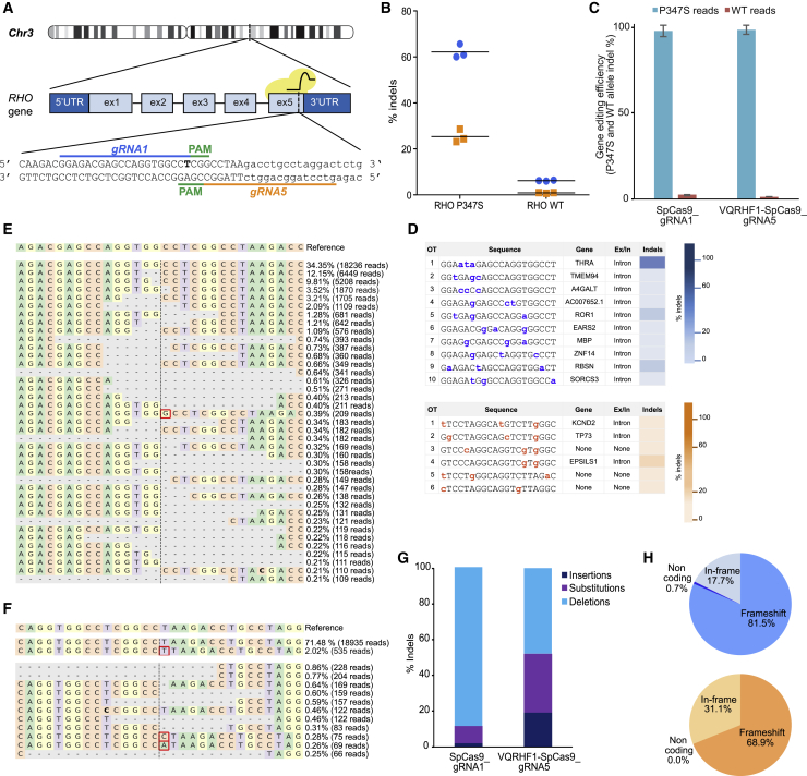
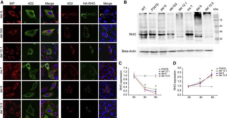
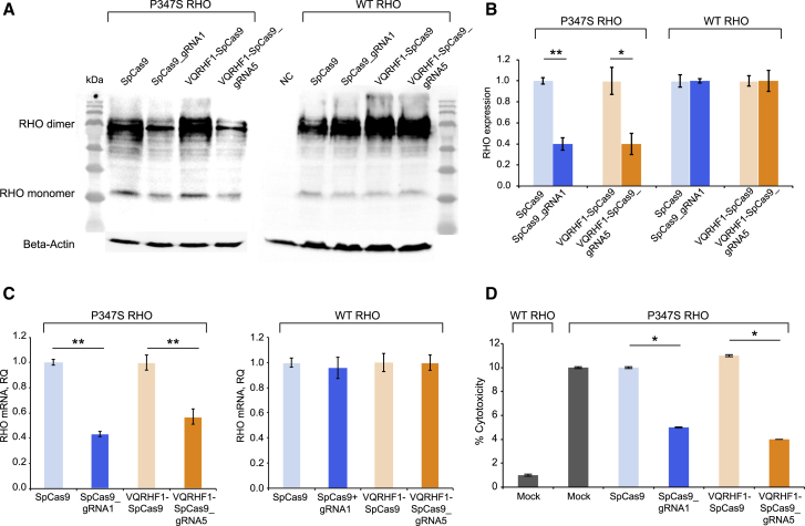
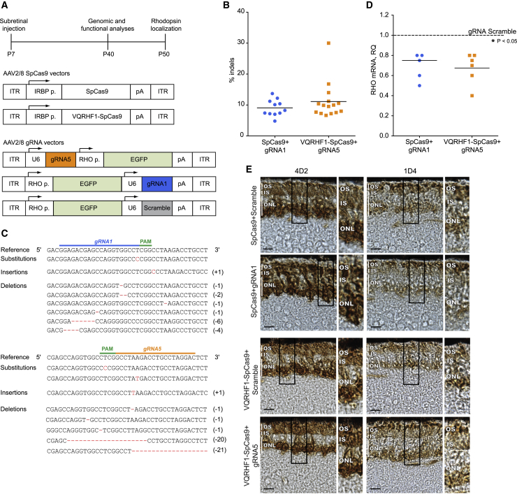
Retinitis pigmentosa (RP) is a group of progressive retinal degenerations of mostly monogenic inheritance, which cause blindness in about 1:3,500 individuals worldwide. Heterozygous variants in the rhodopsin (RHO) gene are the most common cause of autosomal dominant RP (adRP). Among these, missense variants at C-terminal proline 347, such as p.Pro347Ser, cause severe adRP recurrently in European affected individuals. Here, for the first time, we use CRISPR/Cas9 to selectively target the p.Pro347Ser variant while preserving the wild-type RHO allele in vitro and in a mouse model of adRP. Detailed in vitro, genomic, and biochemical characterization of the rhodopsin C-terminal editing demonstrates a safe downregulation of p.Pro347Ser expression leading to partial recovery of photoreceptor function in a transgenic mouse model treated with adeno-associated viral vectors. This study supports the safety and efficacy of CRISPR/Cas9-mediated allele-specific editing and paves the way for a permanent and precise correction of heterozygous variants in dominantly inherited retinal diseases.

Keywords: AAV vector; CRISPR-Cas9 editing; Rhodopsin; retinitis pigmentosa; transgenic mice.


Conflict of interest statement

C.P., M.L., D.B., A.A., and A.R. are listed as inventors on a patent application related to this work.

Figures

Figure 1
Figure 1
CRISPR/Cas9 targeting of c.1039C>T RHO dominant variant encoding p.Pro347Ser (A) Schematic representation of human chromosome 3. The picture illustrates two gRNAs (gRNA1 and gRNA5) targeting the variant (T, in bold) in the exon 5 of RHO and the PAM sequences. Capital letters indicate the exon 5, whereas the 3′ UTR is in lowercase. (B) CRISPResso analysis of NGS data obtained on Pro347Ser and WT RHO HeLa clones transfected with effector plasmids (SpCas9_gRNA1 in blue and VQRHF1-SpCas9_gRNA5 in orange). The experiment was performed in triplicate, and the mean is presented. (C) CRISPResso analysis of indels occurring in c.1039C>T RHO transgene and in the endogenous WT RHO gene after transfection of effector plasmids in Pro347Ser RHO HeLa clone. The experiment was performed in triplicate and is presented as mean ± SEM. (D) Indels analysis of off-target sites predicted for gRNA1 (top) and for gRNA5 (bottom). The color bars (blue and orange) represent values ascending from bottom to top ranking the indels frequency. (E and F) CRISPResso graphic representation of indels scored in the target site of Pro347Ser RHO HeLa cells transfected with SpCas9_gRNA1 (E) and VQRHF1-SpCas9_gRNA5 (F). The top sequence is the unmodified reference. The percentage of indel frequency and the number of reads scored are indicated. Red boxes indicate nucleotide insertion and nucleotide in bold indicates substitution. (G) Type of indels, and their relative percentage, generated in Pro347Ser RHO HeLa clone transfected with SpCas9_gRNA1 and VQRHF1-SpCas9_gRNA5. (H) CRISPResso analysis of indels generated by SpCas9_gRNA1 (top pie chart) and VQRHF1-SpCas9_gRNA5 (bottom pie chart) leading to frameshift or in-frame alterations.
Figure 2
Figure 2
Biochemical characterization of the most frequent RHO variants generated after editing (A) Immunofluorescence analysis of CHO cells transfected with plasmids coding for RHO variants. Permeabilized cells (left) were stained with anti-BIP and anti-4D2 antibodies, and the scale bar represents 5 μm. Cells not permeabilized (right) were stained with anti-4D2 and anti-HA antibodies, and the scale bar represents 10 μm. (B) Immunoblot analysis of WT, p.Pro347Ser, and most frequent RHO variants generated after editing expressed in CHO cells transfected with the respective coding plasmids. Anti-HA antibody was used to detect rhodopsin (RHO). The immunoblotting was normalized with an anti-beta-actin antibody. (C) Densitometric analysis of immunoblots performed on CHO cells transfected with plasmids coding for p.Pro347Ser, del9, del12.1, and del12.5 RHO variants and treated with 10 μg/mL cycloheximide (CHX). The experiment was performed in triplicate and is presented as mean ± SEM. p value < 0.05. (D) Densitometric analysis of immunoblots performed on CHO cells transfected with plasmids coding for p.Pro347Ser, del9, del12.1, and del12.5 RHO variants and treated with 50 μM MG-132 proteasome inhibitor. The experiment was performed in triplicate and is presented as mean ± SEM. p value < 0.05.
Figure 3
Figure 3
Efficient knockdown of p.Pro347Ser RHO expression in vitro (A) Immunoblot for rhodopsin protein expressed in Pro347Ser and WT RHO HeLa clones transfected with SpCas9_gRNA1 and VQRHF1-SpCas9_gRNA5 plasmids and control plasmids (SpCas9 and VQRHF1-SpCas9). 4D2 antibody was used to detect rhodopsin. The immunoblotting was normalized with anti-beta-actin antibody. (B) Densitometric quantification of rhodopsin protein level normalized to beta-actin after editing. The experiment was performed in triplicate and is presented as mean ± SEM. p < 0.05; ∗∗p < 0.01. (C) Pro347Ser and WT RHO HeLa clones were transfected with SpCas9_gRNA1 and VQRHF1-SpCas9_gRNA5 plasmids and control plasmids (SpCas9 and VQRHF1-SpCas9). The relative quantity (RQ) was calculated with the 2-ΔΔCT quantification and is reported in the y axis. Each sample was run in triplicate. ∗∗p < 0.01. (D) Lactate dehydrogenase (LDH) assay of Pro347Ser RHO HeLa clone transfected with SpCas9_gRNA1 and VQRHF1-SpCas9_gRNA5 plasmids and control plasmids (SpCas9 and VQRHF1-SpCas9). Mock-transfected Pro347Ser RHO HeLa cells and WT RHO HeLa cells were used as positive and negative controls. The experiment was performed in triplicate and is presented as mean ± SEM. p value < 0.05.
Figure 4
Figure 4
Allele-specific editing in mouse photoreceptors (A) (Top) a scheme of the experimental timeline is depicted; (middle) AAV2/8 vectors expressing the WT or VQRHF1 SpCas9 under the control of IRBP promoter are schematized; (bottom) AAV2/8 vectors expressing the gRNA (gRNA1, gRNA5, or scramble) and the GFP under the control of RHO promoter. (B) Indels frequency determined by NGS in retinae injected with effector vectors. (C) Representation of indels scored in retinae treated with gRNA1 or gRNA5 AAV vector coupled to appropriate AAV-SpCas9 vector. Nucleotides inserted or deleted are reported on the right. (D) Downregulation of RHO transcript coding for p.Pro347Ser RHO in retinae injected with effector vectors with respect to retinae treated with scramble vectors. The averages are depicted with a bar. p < 0.05. (E) Rhodopsin localization was investigated at P50 in retinae injected with effector vectors using 4D2 and 1D4 antibodies against rhodopsin. Representative images are shown. Scale bar represents 10 μm. Zoomed areas of photoreceptors are shown on the side. OS, outer segment; IS, inner segment; ONL, outer nuclear layer.
Figure 5
Figure 5
Significant improvement of retinal electrical function and pupillary light response (A and B) Pro347Ser transgenic mice injected with effector or control AAV2/8 vectors were examined at P40 by ERG (A), as shown in the data point distribution of B-wave amplitude at 20 cd.s/m2, and PLR analysis (B). Individual eyes are depicted as squares. Data are presented as mean ± SEM. p < 0.05; ∗∗p < 0.01.

References

    1. Verbakel S.K., van Huet R.A.C., Boon C.J.F., den Hollander A.I., Collin R.W.J., Klaver C.C.W., Hoyng C.B., Roepman R., Klevering B.J. Non-syndromic retinitis pigmentosa. Prog. Retin. Eye Res. 2018;66:157–186. - PubMed
    1. Hamel C. Retinitis pigmentosa. Orphanet J. Rare Dis. 2006;1:40. - PMC - PubMed
    1. Parmeggiani F. Clinics, epidemiology and genetics of retinitis pigmentosa. Curr. Genomics. 2011;12:236–237. - PMC - PubMed
    1. Benati D., Patrizi C., Recchia A. Gene editing prospects for treating inherited retinal diseases. J. Med. Genet. 2020;57:437–444. - PubMed
    1. Dias M.F., Joo K., Kemp J.A., Fialho S.L., da Silva Cunha A., Jr., Woo S.J., Kwon Y.J. Molecular genetics and emerging therapies for retinitis pigmentosa: Basic research and clinical perspectives. Prog. Retin. Eye Res. 2018;63:107–131. - PubMed

Publication types

MeSH terms