Review
. 2021 Jan-Jun:296:100336.
doi: 10.1016/j.jbc.2021.100336. Epub 2021 Jan 26.

Maturation, inactivation, and recovery mechanisms of soluble guanylyl cyclase

Affiliations
Review

Maturation, inactivation, and recovery mechanisms of soluble guanylyl cyclase

Dennis J Stuehr et al. J Biol Chem. 2021 Jan-Jun.

Abstract

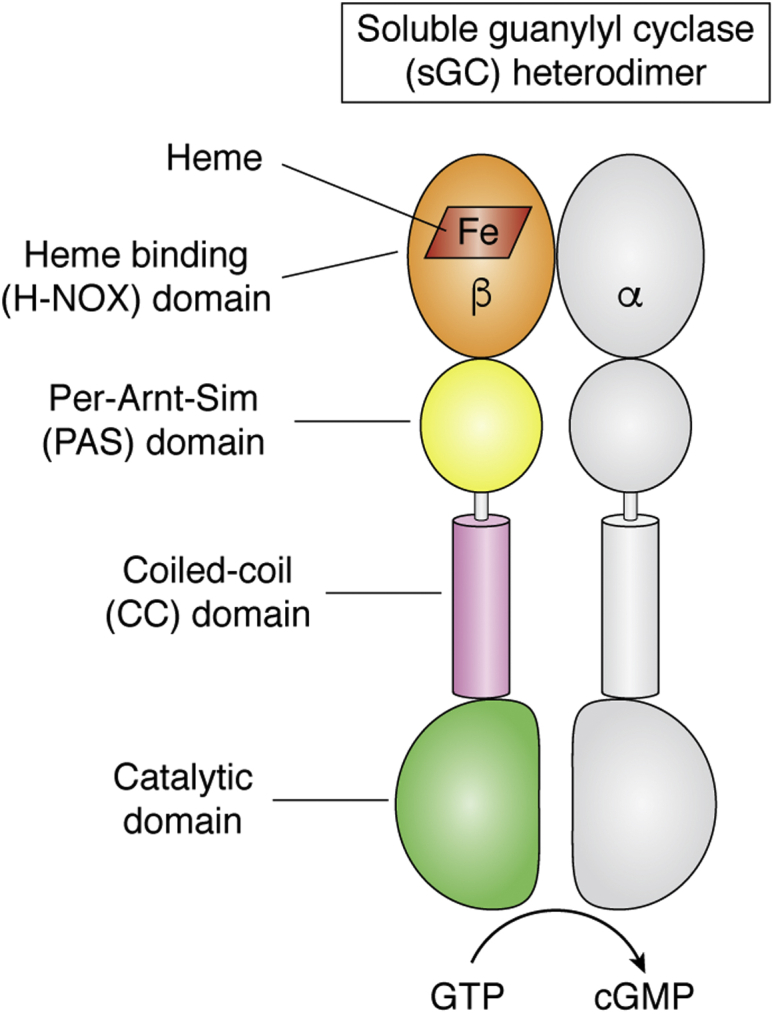
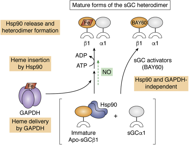
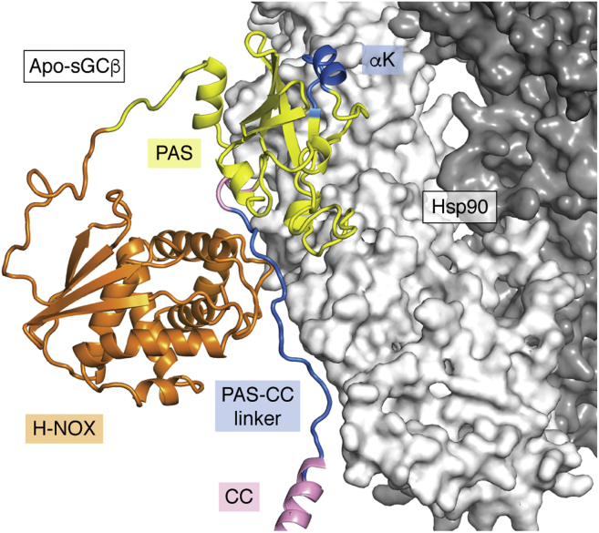
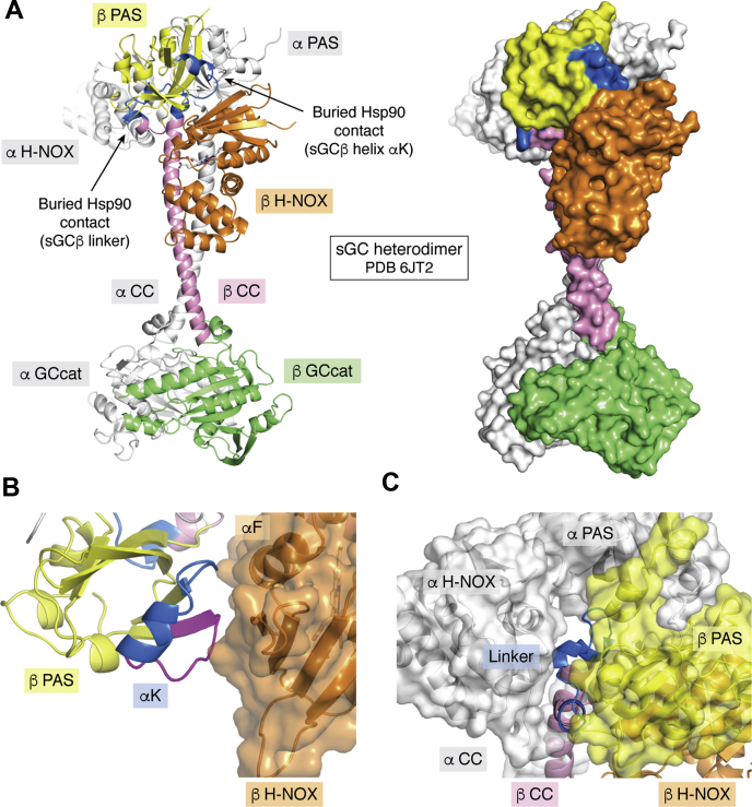
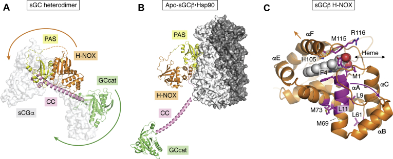
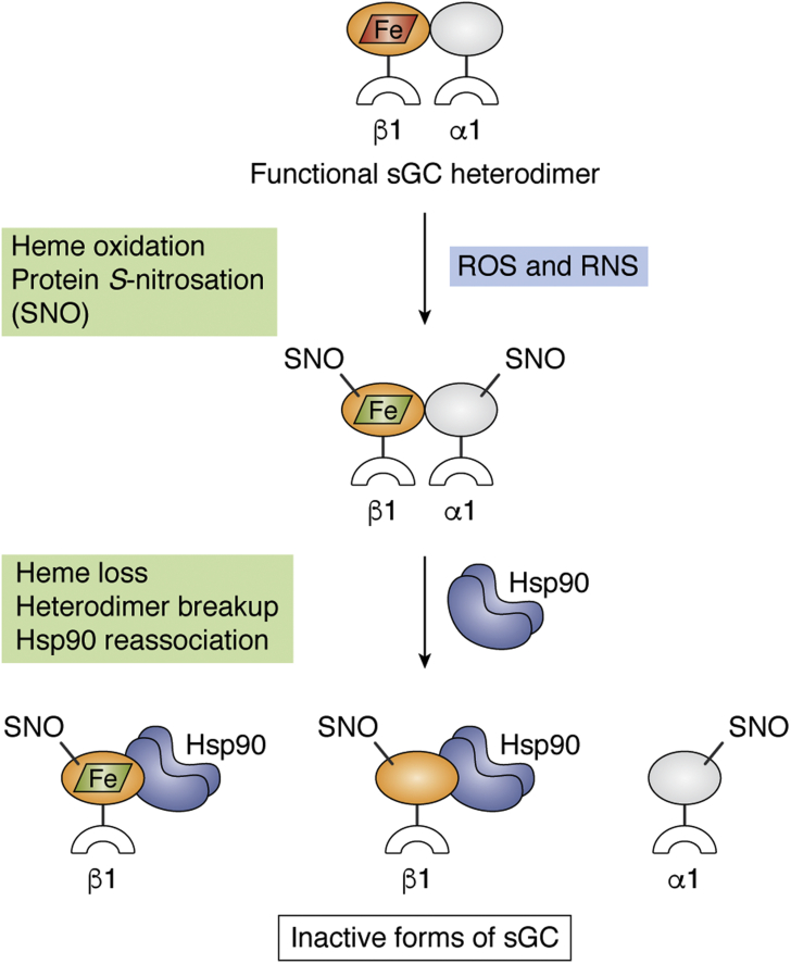
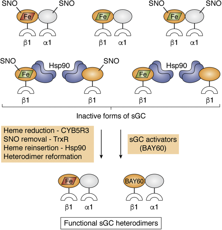
Soluble guanylate cyclase (sGC) is a heme-containing heterodimeric enzyme that generates many molecules of cGMP in response to its ligand nitric oxide (NO); sGC thereby acts as an amplifier in NO-driven biological signaling cascades. Because sGC helps regulate the cardiovascular, neuronal, and gastrointestinal systems through its cGMP production, boosting sGC activity and preventing or reversing sGC inactivation are important therapeutic and pharmacologic goals. Work over the last two decades is uncovering the processes by which sGC matures to become functional, how sGC is inactivated, and how sGC is rescued from damage. A diverse group of small molecules and proteins have been implicated in these processes, including NO itself, reactive oxygen species, cellular heme, cell chaperone Hsp90, and various redox enzymes as well as pharmacologic sGC agonists. This review highlights their participation and provides an update on the processes that enable sGC maturation, drive its inactivation, or assist in its recovery in various settings within the cell, in hopes of reaching a better understanding of how sGC function is regulated in health and disease.

Keywords: Hsp90; cell signaling; cytochrome b5 reductase; hypertension; nitric oxide; protein nitrosation; protein oxidation.


Conflict of interest statement

Conflict of interest The authors declare that they have no conflicts of interest with the contents of this article.

Figures

Figure 1
Figure 1
The sGC heterodimer. The protein domains are noted, and heme is shown as a red parallelogram.
Figure 2
Figure 2
Maturation of the sGC heterodimer. Steps in maturation start from the bottom of the Figure and continue upward. For simplicity, the sGC subunits are depicted as orange or gray ovals representing the H-NOX plus PAS domains attached to a white crescent that represents their CC and catalytic domains. Following translation, the immature apo-sGCβ1 subunit forms a complex with Hsp90 and does not associate with an sGCα1 subunit. GAPDH delivers heme (red parallelogram), which is inserted into the apo-sGCβ1 in an ATP-driven, Hsp90-dependent process. Heme insertion triggers Hsp90 dissociation from sGCβ1, and this allows it to bind an sGCα1 subunit to form the mature sGC heterodimer. Low levels of NO (green) can stimulate the maturation process in an as yet undefined way (green arrow). sGC activator compounds like BAY 60 can bind within immature apo-sGCβ1 and drive maturation to the sGC heterodimer independent of Hsp90, GAPDH, or heme. CC, coiled-coil; PAS, Per-Arnt-Sim; sGC, soluble guanylate cyclase.
Figure 3
Figure 3
Chaperoning of apo-sGCβ by Hsp90. The model of the apo-sGCβ–Hsp90 complex was generated as described (76). The Hsp90 dimer is colored light/dark gray. The H-NOX, PAS, linker, and coiled-coil domains of apo-sGCβ are colored orange, yellow, pink, and pink, respectively, while segments that directly contact Hsp90 as judged by HDX-MS, and other experimental data are colored blue and separately labeled (helix αK and a portion of the PAS-CC linker). CC, coiled-coil; HDX-MS, hydrogen-deuterium exchange mass spectrometry; PAS, Per-Arnt-Sim; sGC, soluble guanylate cyclase.
Figure 4
Figure 4
Structure of the sGC heterodimer shows occlusion of the sGCβ Hsp90-interacting sites.A, the sGC heterodimer in the NO-bound state (PDB ID 6jt2; Kang et al. [17]) is shown in cartoon and surface presentations. The H-NOX, PAS, linker, coiled-coil, and catalytic domains of sGCβ are colored orange, yellow, pink, pink, and green, respectively. The entirety of sGCα is colored white. Segments of the sGCβ PAS and linker domains that interact directly with Hsp90 are colored blue and labeled. B, close-up showing the intradomain packing between the H-NOX domain and the Hsp90 binding segment around helix αK in the heterodimer. A segment near helix αK that exhibits increased solvent exposure when Hsp90 binds to apo-sGCβ is colored magenta. C, close-up showing how the Hsp90 binding segment within the sGCβ PAS-CC linker is buried at the interfaces of the sGCα H-NOX and the PAS and CC domains of sGCβ in the sGC heterodimer. CC, coiled-coil; NO, nitric oxide; PAS, Per-Arnt-Sim; sGC, soluble guanylate cyclase.
Figure 5
Figure 5
Hsp90 stabilizes a structurally open conformation of apo-sGCβ to enable heme insertion.A and B, conformations of the sGCβ subunit when it is in the sGC heterodimer (PDB ID 6jt2; Kang et al. [17]) versus in complex with Hsp90 (docking model, Dai et al. [76]). The yellow PAS domain is kept in an identical orientation in both panels. The arrows indicate movements of sGCβ domains that must occur relative to the yellow PAS domain in order to allow Hsp90 binding, thus creating a more open structure with a free H-NOX domain. C, closeup of the sGCβ H-NOX domain when present in the sGC heterodimer (PDB 6jt0; Kang et al. [17]) with the bound heme shown in sphere representation. The N-terminal αA helix and selected residues colored in magenta undergo deprotection when Hsp90 binds to apo-sGCβ. The magenta and orange arrows indicate potential Hsp90-driven movements of helices αA, αB, αC, and αF away from the rest of the H-NOX domain that increase exposure of the heme pocket to enable heme delivery. The dark gray arrow indicates a potential heme entry pathway. NO, nitric oxide; PAS, Per-Arnt-Sim; sGC, soluble guanylate cyclase.
Figure 6
Figure 6
sGC inactivation pathways and potential structural consequences. When functional sGC heterodimer (top) is exposed to reactive oxygen and/or reactive nitrogen species (ROS and RNS) it can become unresponsive toward NO by undergoing oxidation of its ferrous heme to ferric (red and green parallelograms) and protein modifications like Cys S-nitrosation (SNO). These events alone or in combination may lead to breakup of the sGC heterodimer, heme loss, and/or rebinding of Hsp90 to the freed sGCβ1 subunit. NO, nitric oxide; PAS, Per-Arnt-Sim; sGC, soluble guanylate cyclase.
Figure 7
Figure 7
Inactivated forms of sGC and potential recovery pathways. Inactivated sGC may exist in at least seven forms in cells (top): A ferrous heme-containing (red parallelogram) heterodimer with Cys-NO (SNO) modifications, a ferric heme-containing (green parallelogram) heterodimer with or without SNO modifications, and dissociated forms in which a SNO-modified or SNO-free sGCβ subunit that either does or does not contain ferric heme is in complex with Hsp90. Conversion of the various inactive sGC forms to a functional sGC heterodimer may involve the steps and proteins and the small molecules noted in the colored boxes. sGC, soluble guanylate cyclase.

Similar articles

Cited by

References

    1. Garthwaite J. New insight into the functioning of nitric oxide-receptive guanylyl cyclase: Physiological and pharmacological implications. Mol. Cell Biochem. 2010;334:221–232. - PubMed
    1. Moncada S., Higgs E.A. The discovery of nitric oxide and its role in vascular biology. Br. J. Pharmacol. 2006;147 Suppl 1:S193–201. - PMC - PubMed
    1. Michel T., Vanhoutte P.M. Cellular signaling and NO production. Pflugers Arch. 2010;459:807–816. - PMC - PubMed
    1. Buys E., Sips P. New insights into the role of soluble guanylate cyclase in blood pressure regulation. Curr. Opin. Nephrol. Hypertens. 2014;23:135–142. - PMC - PubMed
    1. Pyriochou A., Papapetropoulos A. Soluble guanylyl cyclase: More secrets revealed. Cell Signal. 2005;17:407–413. - PubMed

Publication types

LinkOut - more resources