. 2021:29:102571.
doi: 10.1016/j.nicl.2021.102571. Epub 2021 Jan 19.

Homotopic redistribution of functional connectivity in insula-centered diffuse low-grade glioma

Affiliations

Homotopic redistribution of functional connectivity in insula-centered diffuse low-grade glioma

Fabien Almairac et al. Neuroimage Clin. 2021.

Abstract

Objective: In the event of neural injury, the homologous contralateral brain areas may play a compensatory role to avoid or limit the functional loss. However, this dynamic strategy of functional redistribution is not clearly established, especially in the pathophysiological context of diffuse low-grade glioma. Our aim here was to assess the extent to which unilateral tumor infiltration of the insula dynamically modulates the functional connectivity of the contralesional one.

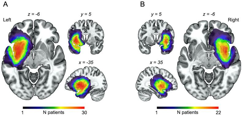
Methods: Using resting-state functional connectivity MRI, a seed-to-ROI approach was employed in 52 insula-centered glioma patients (n = 30 left and 22 right) compared with 19 age-matched healthy controls.

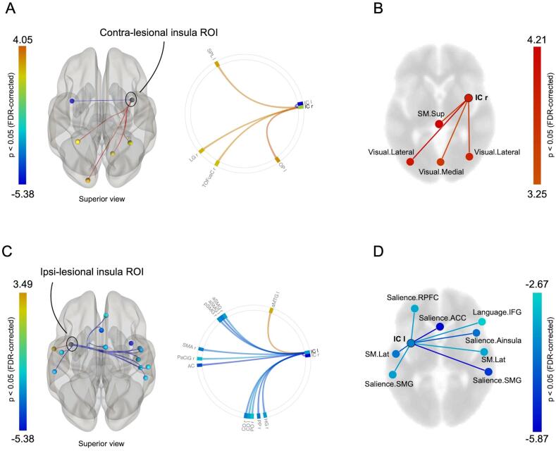
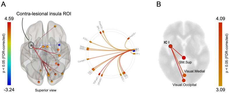
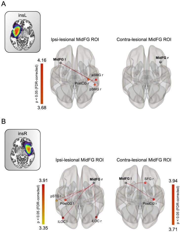
Results: Unsurprisingly, a significant decrease of the inter-insular connectivity was observed in both patient groups. More importantly, the analyses revealed a significant increase of the contralesional insular connectivity towards both cerebral hemispheres, especially in cortical areas forming the visual and the sensorimotor networks. This functional redistribution was not identified when the analyses were performed on three control regions for which the homologous area was not impaired by the tumor. This overall pattern of results indicates that massive infiltration of the insular cortex causes a significant redeployment of the contralesional functional connectivity.

Conclusion: This general finding suggests that the undamaged insula plays a role in the functional compensation usually observed in this patient population, and thus provides compelling support for the concept of homotopic functional plasticity in brain-damaged patients.

Keywords: Functional MRI; Functional connectivity; Glioma; Homotopic; Insula; Plasticity.


Conflict of interest statement

The authors declare that they have no known competing financial interests or personal relationships that could have appeared to influence the work reported in this paper.

Figures

Fig. 1
Fig. 1
Lesion overlap map for (A) left insular glioma and (B) right insular glioma patients. As expected, the maximum overlap occurred in the insula.
Fig. 2
Fig. 2
Tumor-related modulation of insular connectivity in left insular glioma patients versus healthy controls. (A) Contralesional insula seed-to-ROI map and connectome ring. (B) Contralesional seed-to-network map displayed on an axial slide. (C) Ipsilesional insula seed-to-ROI map and connectome ring. (D) Ipsilesional insula seed-to-network map displayed on an axial slide. Only significant T-values (p < 0.05 FDR-corrected) are displayed. ACC = anterior cingulate cortex; IC = insular cortex; IFG = inferior frontal gyrus; FDR = false discovery rate; RPFC = rostral prefrontal cortex; SM.Lat = lateral sensorimotor; SM.Sup = superior sensorimotor; SMG = supramarginal gyrus.
Fig. 3
Fig. 3
Tumor-related modulation of insular connectivity in right insular glioma patients versus healthy controls. (A) Contralesional seed-to-ROI map and connectome ring. (B) Contralesional seed-to-network map displayed on an axial slide. Only significant T-values (p < 0.05 FDR-corrected) are displayed. IC = insular cortex; SM.Sup = superior sensorimotor.
Fig. 4
Fig. 4
Modulation of the homologous MidFG connectivity in left and right insular glioma patients versus healthy controls. (A) Ipsi- and contralesional MidFG seed-to-ROI map for left insular glioma patient. (B) Contra- and Ipsilesional seed-to-ROI map for right insular glioma. Only significant T-values (p < 0.05 FDR-corrected) are displayed. InsL = left insular glioma patients; InsR = right insular glioma patients; iLOC = inferior lateral occipital; MidFG = middle frontal gyrus; SMG = supramarginal gyrus; postCG = posterior central gyrus; SFG = superior frontal gyrus; STG = superior temporal gyrus.

Similar articles

Cited by

References

    1. Agarwal S., Sair H.I., Yahyavi-Firouz-Abadi N., Airan R., Pillai J.J. Neurovascular uncoupling in resting state fMRI demonstrated in patients with primary brain gliomas. J. Magn. Reson. Imaging. 2016;43(3):620–626. doi: 10.1002/jmri.25012. - DOI - PubMed
    1. Almairac F., Duffau H., Herbet G. Contralesional macrostructural plasticity of the insular cortex in patients with glioma: A VBM study. Neurology. 2018;91(20):e1902–e1908. doi: 10.1212/WNL.0000000000006517. - DOI - PubMed
    1. Almairac F., Herbet G., Moritz-Gasser S., de Champfleur N.M., Duffau H. The left inferior fronto-occipital fasciculus subserves language semantics: a multilevel lesion study. Brain Struct. Funct. 2015;220(4):1983–1995. doi: 10.1007/s00429-014-0773-1. - DOI - PubMed
    1. Behzadi Y., Restom K., Liau J., Liu T.T. A component based noise correction method (CompCor) for BOLD and perfusion based fMRI. Neuroimage. 2007;37(1):90–101. doi: 10.1016/j.neuroimage.2007.04.042. - DOI - PMC - PubMed
    1. Boroojerdi B., Diefenbach K., Ferbert A. Transcallosal inhibition in cortical and subcortical cerebral vascular lesions. J. Neurol. Sci. 1996;144(1-2):160–170. doi: 10.1016/S0022-510X(96)00222-5. - DOI - PubMed

LinkOut - more resources