

Multi-factorial considerations for intra-thoracic lymph node evaluations of healthy cats on computed tomographic images
- PMID: 33509167
- PMCID: PMC7844987
- DOI: 10.1186/s12917-021-02771-7
Multi-factorial considerations for intra-thoracic lymph node evaluations of healthy cats on computed tomographic images
Abstract
Background: It is difficult to examine mild to moderate feline intra-thoracic lymphadenopathy via and thoracic radiography. Despite previous information from computed tomographic (CT) images of intra-thoracic lymph nodes, some factors from animals and CT setting were less elucidated. Therefore, this study aimed to investigate the effect of internal factors from animals and external factors from the CT procedure on the feasibility to detect the intra-thoracic lymph nodes. Twenty-four, client-owned, clinically healthy cats were categorized into three groups according to age. They underwent pre- and post-contrast enhanced CT for whole thorax followed by inter-group evaluation and comparison of sternal, cranial mediastinal, and tracheobronchial lymph nodes.
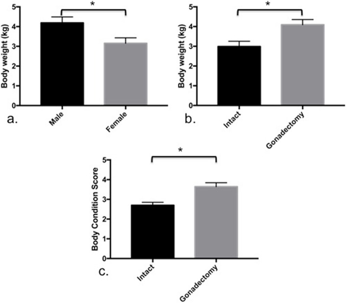
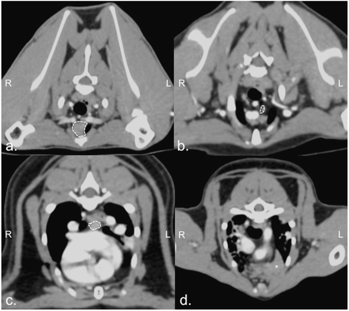
Results: Post contrast-enhanced CT appearances revealed that intra-thoracic lymph nodes of kittens were invisible, whereas the sternal, cranial mediastinal, and tracheobronchial nodes of cats aged over 7 months old were detected (6/24, 9/24 and 7/24, respectively). Maximum width of these lymph nodes were 3.93 ± 0.74 mm, 4.02 ± 0.65 mm, and 3.51 ± 0.62 mm, respectively. By age, lymph node sizes of these cats were not significantly different. Transverse lymph node width of males was larger than that of females (P = 0.0425). Besides, the detection score of lymph nodes was affected by slice thickness (P < 0.01) and lymph node width (P = 0.0049). Furthermore, an irregular, soft tissue structure, possibly the thymus, was detected in all juvenile cats and three mature cats.
Conclusions: Despite additional information on intra-thoracic lymph nodes in CT images, which can be used to investigate lymphatic-related abnormalities, age, sex, and slice thickness of CT images must be also considered.
Keywords: Cat; Computed tomography; Lymph node; Slice thickness; Thorax; Thymus.
Conflict of interest statement
The authors declare that they have no competing interests and do not have any potential conflict of interest to declare.
Figures

References
-
- Aspinall V, O’Reilly M. The blood vascular system. In: O’reilly M, Aspinall V, editors. Introduction to veterinary anatomy and physiology. 1. Toronto: Butterworth Heinemann; 2004. pp. 94–97.
-
- Sisson S, Grossman JD, Getty R. Lymphatic system. In: Rosenbaum CE, Ghoshal NG, Hillman D, editors. Sisson and Grossman’s the anatomy of the domestic animals. Philadelphia: Saunders; 1975. pp. 1652–1670.
-
- Vollerhaus B. Lymphatic system. In: Schummer A, Wilkens H, Vollmerhaus B, Habermehl KH, editors. The anatomy of the domestic animals. Germany: Springer; 1981. pp. 357–359.
MeSH terms
Grants and funding
LinkOut - more resources
Full Text Sources
Other Literature Sources
Medical
Miscellaneous
