

Epigenetic immunomodulatory effect of eugenol and astaxanthin on doxorubicin cytotoxicity in hormonal positive breast Cancer cells
- PMID: 33509300
- PMCID: PMC7842008
- DOI: 10.1186/s40360-021-00473-2
Epigenetic immunomodulatory effect of eugenol and astaxanthin on doxorubicin cytotoxicity in hormonal positive breast Cancer cells
Abstract
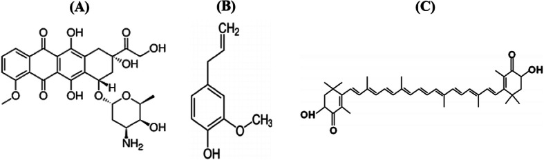
Background: Hormonal receptor positive (HR+) breast cancer is the most commonly diagnosed molecular subtype of breast cancer; which showed good response to doxorubicin (DOX)-based chemotherapy. Eugenol (EUG) and astaxanthin (AST) are natural compounds with proved epigenetic and immunomodulatory effects in several cancer cell lines. This study has been initiated to investigate the molecular mechanism (s) whereby EUG and AST could enhance DOX cytotoxicity in MCF7 cells.
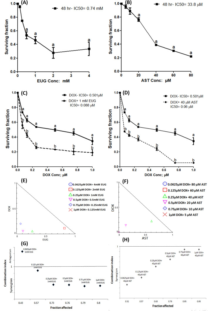
Methods: Cytotoxic activity of DOX alone and combined with either 1 mM EUG or 40 μM AST was performed using sulphorhodamine-B assay in MCF7 cells. Global histones acetylation and some immunological markers were investigated using ELISA, western blotting and quantitative RT-PCR techniques. Functional assay of multidrug resistance was performed using rhodamine 123 and Hoechst 3342 dyes. Flow cytometry with annexin V and propidium iodide were used to assess the change in cell cycle and apoptosis along with the expression of some differentiation, apoptosis and autophagy proteins.
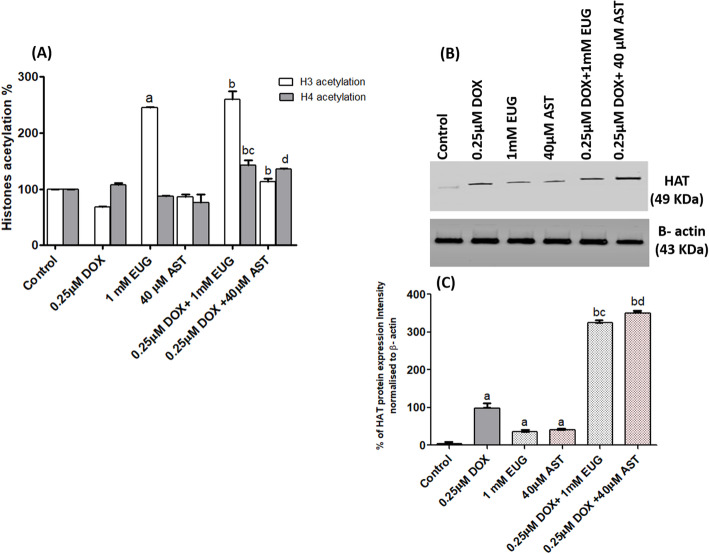
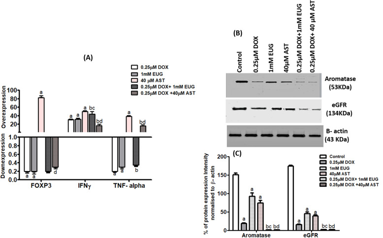
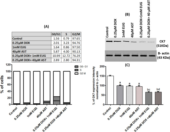
Results: DOX alone resulted in concentration-dependent cytotoxicity with IC50 of 0.5 μM. Both EUG and AST significantly increased DOX cytotoxicity which is manifested as a significant decrease in DOX IC50 from 0.5 μM to 0.088 μM with EUG and to 0.06 μM with AST. Combinations of DOX with 1 mM EUG or 40 μM AST significantly increased the level of histones acetylation and histone acetyl transferase expression, while reduced the expression of aromatase and epidermal growth factor receptor (EGFR) when compared with 0.25 μM DOX alone. Also both combinations showed higher uptake of rhodamine but lower of Hoechst stains, along with increased the percentage of caspase 3, and decreased the expression of CK7 and LC3BI/II ratio. EUG combination induced IFγ but reduced TNFα causing shifting of cells from G2/M to S and G0/ G1 phases. Combination of DOX with EUG induced apoptosis through the higher BAX/ BCl2 ratio, while with AST was through the increase in caspase 8 expressions.
Conclusion: EUG and AST potentiated the anticancer activity of DOX through epigenetic histones acetylation along with the immunonomodulation of different apoptotic approaches in MCF7 cells.
Keywords: Astaxanthin; Breast Cancer cells; Doxorubicin; Eugenol.
Conflict of interest statement
The authors declared that have no competing interest.
Figures


Similar articles
-
Low-dose doxorubicin with carotenoids selectively alters redox status and upregulates oxidative stress-mediated apoptosis in breast cancer cells.Food Chem Toxicol. 2018 Aug;118:675-690. doi: 10.1016/j.fct.2018.06.027. Epub 2018 Jun 18. Food Chem Toxicol. 2018. PMID: 29920287
-
Metabolomic Identification of Anticancer Metabolites of Australian Propolis and Proteomic Elucidation of Its Synergistic Mechanisms with Doxorubicin in the MCF7 Cells.Int J Mol Sci. 2021 Jul 22;22(15):7840. doi: 10.3390/ijms22157840. Int J Mol Sci. 2021. PMID: 34360606 Free PMC article.
-
Breviscapine reverses doxorubicin resistance in breast cancer and its related mechanisms.Thorac Cancer. 2023 Sep;14(27):2785-2792. doi: 10.1111/1759-7714.15072. Epub 2023 Aug 16. Thorac Cancer. 2023. PMID: 37584258 Free PMC article.
-
Epigenetic modulation of doxorubicin resistance and strategies for enhancing chemotherapeutic sensitivity.Int Rev Cell Mol Biol. 2025;390:186-198. doi: 10.1016/bs.ircmb.2024.09.004. Epub 2024 Oct 7. Int Rev Cell Mol Biol. 2025. PMID: 39864895 Review.
-
A review of six bioactive compounds from preclinical studies as potential breast cancer inhibitors.Mol Biol Rep. 2025 Feb 5;52(1):203. doi: 10.1007/s11033-025-10300-0. Mol Biol Rep. 2025. PMID: 39907697 Review.
Cited by
-
Molecular Mechanisms of Action of Eugenol in Cancer: Recent Trends and Advancement.Life (Basel). 2022 Nov 6;12(11):1795. doi: 10.3390/life12111795. Life (Basel). 2022. PMID: 36362950 Free PMC article. Review.
-
From Natural Sources to Synthetic Derivatives: The Allyl Motif as a Powerful Tool for Fragment-Based Design in Cancer Treatment.J Med Chem. 2023 Mar 23;66(6):3703-3731. doi: 10.1021/acs.jmedchem.2c01406. Epub 2023 Mar 1. J Med Chem. 2023. PMID: 36858050 Free PMC article. Review.
-
Combination of Conventional Drugs with Biocompounds Derived from Cinnamic Acid: A Promising Option for Breast Cancer Therapy.Biomedicines. 2023 Jan 19;11(2):275. doi: 10.3390/biomedicines11020275. Biomedicines. 2023. PMID: 36830811 Free PMC article. Review.
-
Eugenol: An Insight Into the Anticancer Perspective and Pharmacological Aspects.Food Sci Nutr. 2025 Aug 3;13(8):e70727. doi: 10.1002/fsn3.70727. eCollection 2025 Aug. Food Sci Nutr. 2025. PMID: 40761498 Free PMC article. Review.
-
Wild epiphytic Bangladeshi orchids Cymbidium aloifolium (L.) Sw. and Papilionanthe teres (Roxb.) Lindl. potentially modulates the immune functions in Swiss albino mice.J Adv Vet Anim Res. 2021 Sep 20;8(3):479-488. doi: 10.5455/javar.2021.h537. eCollection 2021 Sep. J Adv Vet Anim Res. 2021. PMID: 34722747 Free PMC article.
References
-
- American Cancer Society . Breast Cancer Facts & Figures 2019-2020. Atlanta: American Cancer society, Inc.; 2019.
Publication types
MeSH terms
Substances
LinkOut - more resources
Full Text Sources
Other Literature Sources
Medical
Research Materials
Miscellaneous
