Review
. 2021 Aug;46(8):630-639.
doi: 10.1016/j.tibs.2020.12.013. Epub 2021 Jan 25.

ER-Phagy, ER Homeostasis, and ER Quality Control: Implications for Disease

Affiliations
Review

ER-Phagy, ER Homeostasis, and ER Quality Control: Implications for Disease

Susan Ferro-Novick et al. Trends Biochem Sci. 2021 Aug.

Abstract

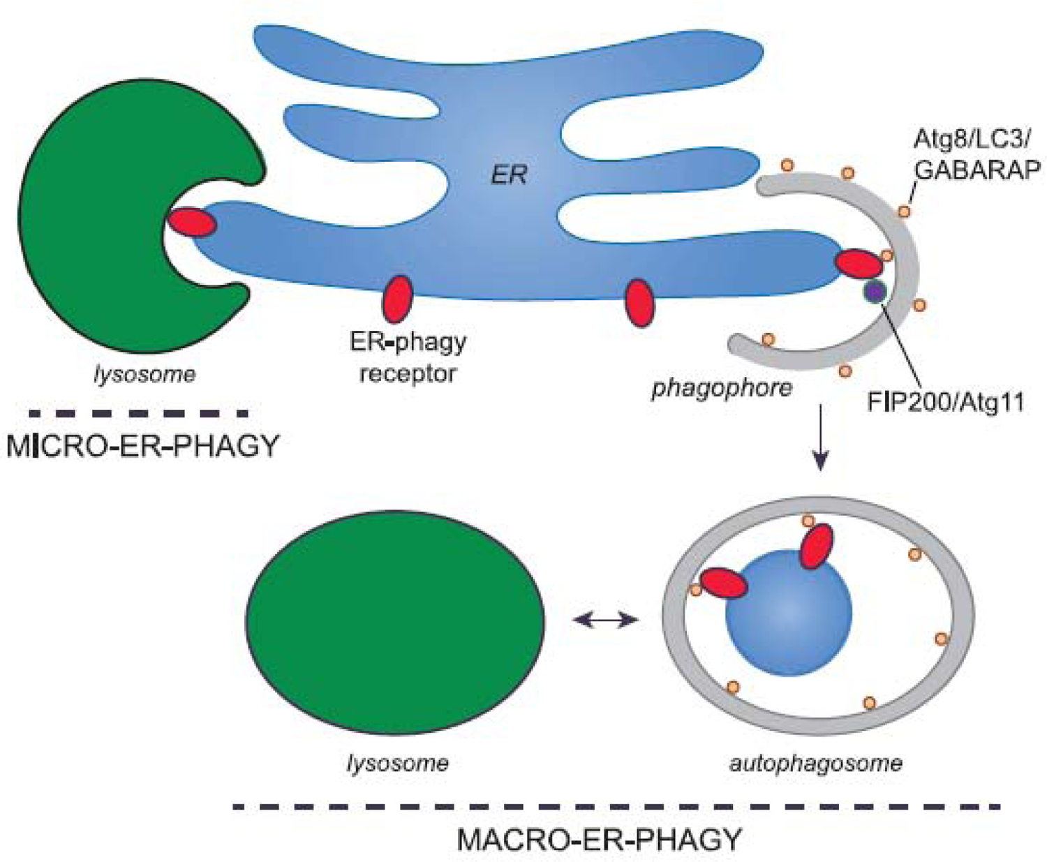
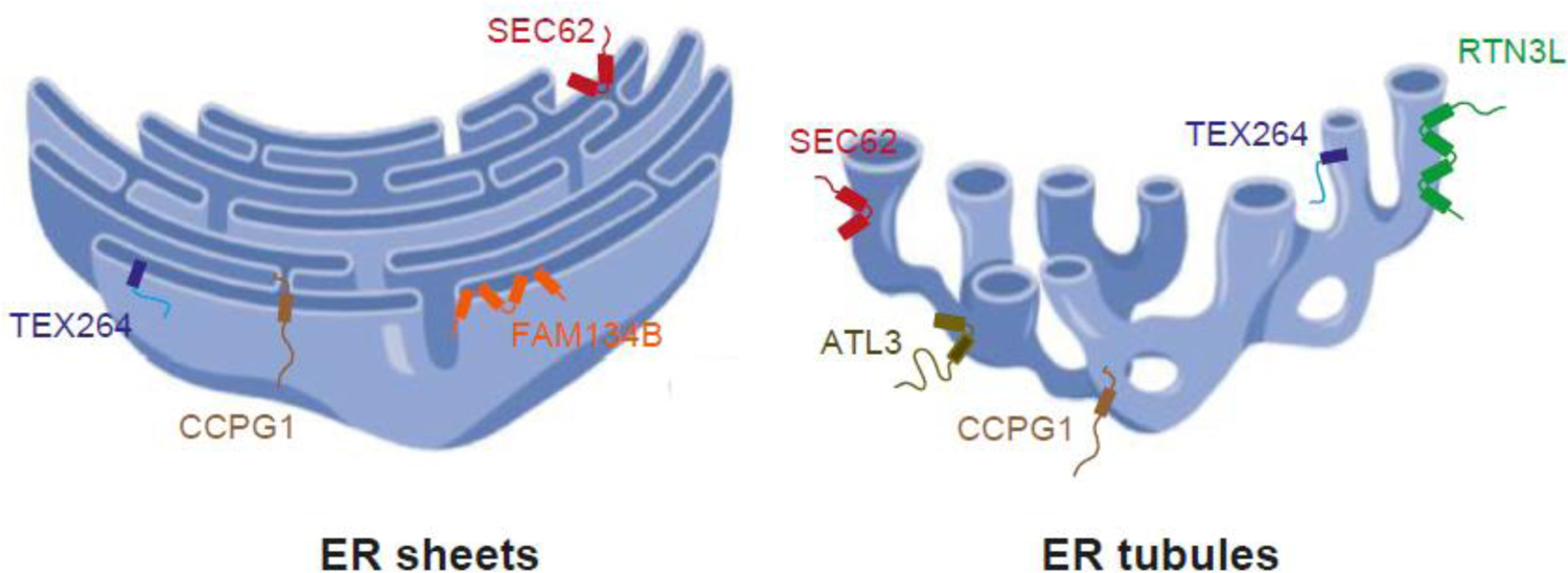
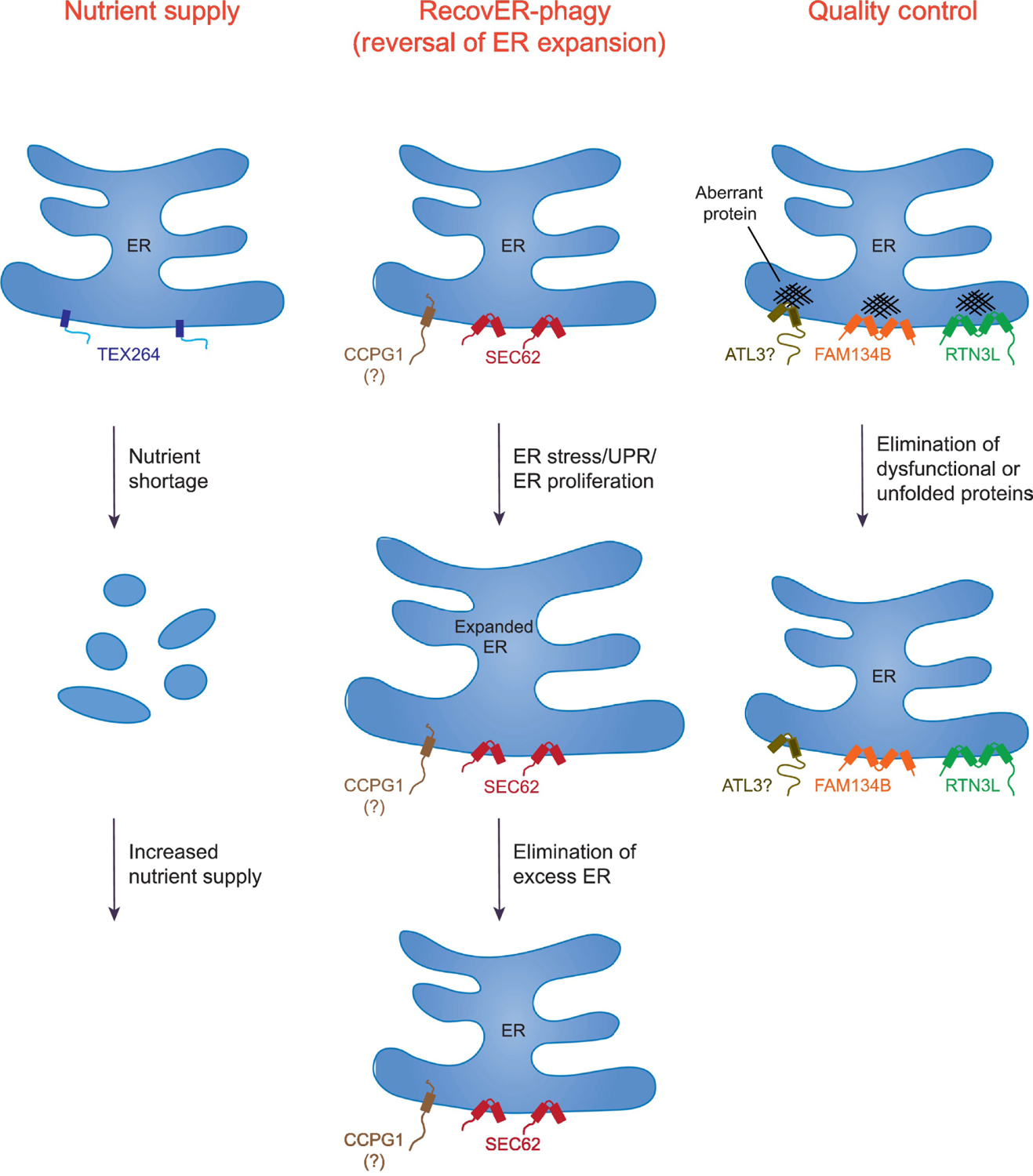
Lysosomal degradation of endoplasmic reticulum (ER) fragments by autophagy, termed ER-phagy or reticulophagy, occurs under normal as well as stress conditions. The recent discovery of multiple ER-phagy receptors has stimulated studies on the roles of ER-phagy. We discuss how the ER-phagy receptors and the cellular components that work with these receptors mediate two important functions: ER homeostasis and ER quality control. We highlight that ER-phagy plays an important role in alleviating ER expansion induced by ER stress, and acts as an alternative disposal pathway for misfolded proteins. We suggest that the latter function explains the emerging connection between ER-phagy and disease. Additional ER-phagy-associated functions and important unanswered questions are also discussed.

Keywords: autophagy receptor; endoplasmic reticulum; human disease; macro-ER-phagy; micro-ER-phagy; proteostasis; reticulophagy.


Conflict of interest statement

Declaration of Interests The authors declare no conflicts of interest.

Figures

Figure 1.
Figure 1.. ER-phagy uses autophagy receptors to package ER into autophagosomes.
During macro-ER-phagy, ER-phagy receptors bind Atg8 in yeast, or LC3 and GABARAP proteins in mammals (tan spheres), to package ER fragments into a phagophore (grey). Some of the ER-phagy receptors also bind the autophagy-inducing ULK complex via its subunit FIP200 (or Atg11 in yeast) to coordinate the autophagy machinery with cargo sequestration. The phagophore expands and then seals to form an autophagosome that delivers ER fragments to the vacuole (yeast) or lysosome (mammals; green) for degradation. During micro-ERphagy, ER fragments are directly engulfed by lysosomes/vacuoles. While in some cases this process involves ER-phagy receptors and LC3 or GABARAP proteins (e.g. during recovER-phagy (9)), micro-ER-phagy in general does not appear to require these factors (e.g. during micro-ER-phagy induced by tunicamycin treatment in yeast (41)).
Figure 2.
Figure 2.. ER-phagy receptors reside in different ER subdomains.
A cross section of an area of sheets (left) and tubules (right) is shown to illustrate the localization of the 6 known mammalian ER membrane ER-phagy receptors. RTN3L localizes to tubules, ATL3 is present at the three-way junctions of the ER tubules, and FAM134B resides on the curved edges of the sheets. The other receptors, i.e., SEC62, CCPG1 and TEX264, are found on flat ER sheets and tubules. Figure created in Biorender.
Figure 3.
Figure 3.. Cellular roles of ER-phagy and the contributions of metazoan ER-phagy receptors.
Shown are the general pathways in which ER-phagy is utilized and the contributing receptors. These pathways include: Nutrient Supply (TEX264), RecovER-phagy (Reversal of ER Expansion) (SEC62 and possibly CCPG1), and Quality Control (FAM134B, RTN3L, and possibly ATL3). Not all the listed pathways are limited to the indicated ER-phagy receptors. For example, while TEX264 contributes to ~50% of autophagic flux during amino acid deprivation, other ER receptors, such as FAM134B, can also participate in starvation-induced ER-phagy, albeit far less efficiently. Note that a significant fraction of the ER is fragmented in the Nutrient Supply pathway to illustrate that TEX264 promotes degradation of a large portion of this organelle during amino acid starvation. SEC62 mediates RecovER-phagy. Although CCPG1 function appears to be regulated by the unfolded protein response (UPR), it remains to be determined whether it also definitively participates in RecovER-phagy. In Quality Control, FAM134B facilitates the removal and degradation of misfolded procollagen (shown) and disease-causing mutant NPC1, whereas misfolded pro-insulin (Akita), proopiomelanocortin (POMC) and arginine vasopressin (pro)-AVP utilize RTN3L. There are no known misfolded proteins that specifically utilize ATL3. Also note that FAM134B and RTN3L are inserted into the lipid bilayer via reticulon domains, whereas the other receptors shown possess bona fide transmembrane segments.
Figure 4.
Figure 4.. Macro-ER-phagy in yeast.
Yeast Atg40 has a domain structure similar to mammalian FAM134B, yet it localizes to the tubular ER, like RTN3L. In yeast, the sheets and tubules in the cytoplasm, and at the cell cortex, are referred to as the cortical ER. Atg40 is largely present on the cortical ER, while Atg39 is principally localized in the nuclear envelope. Atg40, which contains a reticulon-like domain, recruits Atg8 via a LIR motif to initiate cortical macro-ER-phagy. Autophagosome-driven fragmentation of the cortical ER occurs at ER-phagy sites (ERPHS) with the aid of the Lst1-Sec23 complex. In contrast, transport vesicles bud from the ER at ER exit sites (ERES), where the COPII coat complex is assembled. Soluble secretory cargo proteins are loaded into vesicles via a receptor-mediated process, while transmembrane cargo proteins interact with the COPII coat subunits or specific adaptors.

References

    1. Chen S, Novick P, Ferro-Novick S, ER structure and function. Curr Opin Cell Biol 25, 428–433 (2013). - PMC - PubMed
    1. Sun Z, Brodsky JL, Protein quality control in the secretory pathway. J Cell Biol 218, 3171–3187 (2019). - PMC - PubMed
    1. Forrester A et al., A selective ER-phagy exerts procollagen quality control via a Calnexin-FAM134B complex. EMBO J 38, (2019). - PMC - PubMed
    1. Cui Y et al., A COPII subunit acts with an autophagy receptor to target endoplasmic reticulum for degradation. Science 365, 53–60 (2019). - PMC - PubMed
    1. Chino H, Mizushima N, ER-phagy: Quality control and turnover of endoplasmic reticulum. Trends Cell Biol 30, 384–398 (2020). - PubMed

Publication types

Substances

LinkOut - more resources