. 2021 Mar;34(2):160-166.
doi: 10.1016/j.aucc.2020.11.007. Epub 2020 Dec 9.

Complex and prolonged hypercoagulability in coronavirus disease 2019 intensive care unit patients: A thromboelastographic study

Affiliations

Complex and prolonged hypercoagulability in coronavirus disease 2019 intensive care unit patients: A thromboelastographic study

Pierre-Yves Cordier et al. Aust Crit Care. 2021 Mar.

Abstract

Background: A high number of thrombotic complications have been reported in critically ill patients with coronavirus disease 2019 (COVID-19) and appear to be related to a hypercoagulable state. Evidence regarding detection, management, and monitoring of COVID-19-associated coagulopathy is still missing. We propose to describe the thrombus viscoelastic properties to investigate the mechanisms of hypercoagulability in patients with COVID-19.

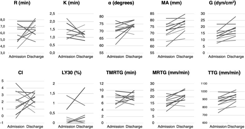
Methods: Thromboelastography (TEG) was performed in 24 consecutive patients admitted to a single intensive care unit for COVID-19 pneumonia, and 10 had a second TEG before being discharged alive from the intensive care unit.

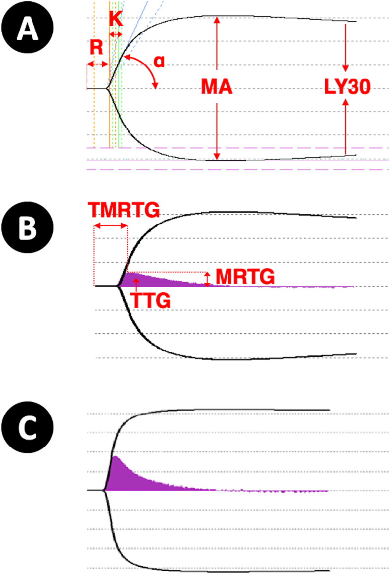
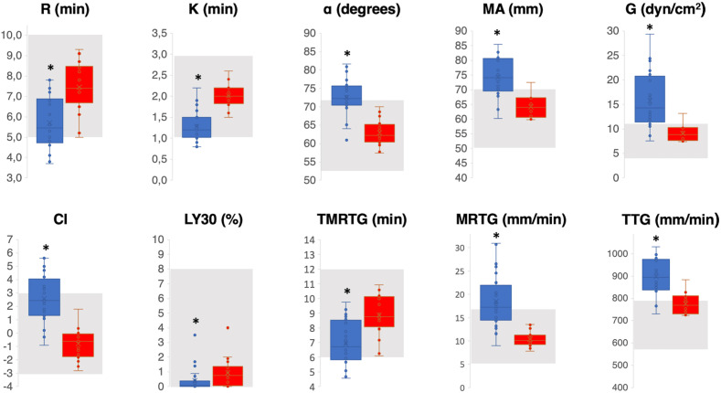
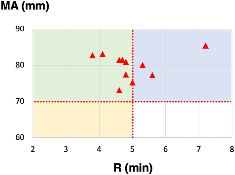
Results: Compared with a group of 20 healthy participants, patients with COVID-19 had significantly decreased values of reaction time, coagulation time, and lysis index and increased values of α angle, maximum amplitude, clot strength, and coagulation index. Velocity curves were consistent with increased generation of thrombin. These values persisted in surviving patients despite their good clinical course.

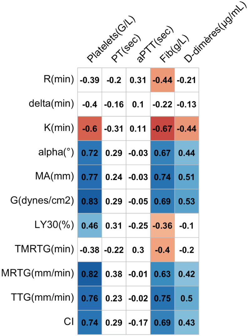
Discussion: In patients with COVID-19, TEG demonstrates a complex and prolonged hypercoagulable state including fast initiation of coagulation and clot reinforcement, low fibrinolysis, high potential of thrombin generation, and high fibrinogen and platelet contribution. The antithrombotic strategy in patients with COVID-19 during intensive care hospitalisation and after discharge should be investigated in further studies.

Keywords: COVID-19; Coagulopathy; Hypercoagulability; Thromboelastography; Thrombosis.


Conflict of interest statement

Conflict of interest On behalf of all the authors, the corresponding author states that there is no conflict of interest.

Figures

Fig. 1
Fig. 1
(A) Main parameters on the TEG trace: R (reaction time), K (coagulation time), α angle, MA (maximum amplitude), and LY30 (lysis index). (B) The velocity curve (V-curve) is the first derivative of the standard TEG trace. Featured parameters are MRTG (maximum rate of thrombus generation) and TMRTG (time to maximum rate of thrombus generation), and the area under the curve gives the TTG (total thrombus generation). (C) Hypercoagulability is characterised by decreased values of R, K, LY30, and TMRTG and increased values of α, MA, MRTG, and TTG. TEG, thromboelastography; R, reaction time; K, coagulation time; MA, maximum amplitude.
Fig. 2
Fig. 2
Patients' TEG values (blue box plot) compared with the reference values established in our laboratory on a control group (red box plot). The grey area indicates the normal value range defined by the manufacturer. An asterisk indicates a statistically significant difference between the two groups. R, reaction time; K, coagulation time; MA, maximum amplitude; CI, coagulation index; TMRTG, time to maximum rate of thrombus generation; MRTG, maximum rate of thrombus generation; TTG, total thrombus generation; TEG, thromboelastography. (For interpretation of the references to colour in this figure legend, the reader is referred to the Web version of this article.)
Fig. 3
Fig. 3
Eleven patients had hypercoagulability defined by CI >3, that could be classified into enzymatic hypercoagulability (R ≤ 5 min, MA ≤70 mm, yellow area), platelet hypercoagulability (R > 5 min, MA >70 mm, blue area), and mixed hypercoagulability (R < 5 min, MA >70 mm, green area). CI, coagulation index; R, reaction time; MA, maximum amplitude. (For interpretation of the references to colour in this figure legend, the reader is referred to the Web version of this article.)
Fig. 4
Fig. 4
Correlation matrix between TEG parameters and standard coagulation tests in 24 patients with COVID-19. Positive correlations are indicated in a blue cell, and negative correlations are indicated in a red cell. Correlation coefficients on a white field are not statistically significant. PT, prothrombin time; aPTT, activated partial thromboplastin time; MA, maximum amplitude; TMRTG, time to maximum rate of thrombus generation; TTG, total thrombus generation; MRTG, maximum rate of thrombus generation; CI, coagulation index; TEG, thromboelastography; COVID-19, coronavirus disease 2019.
Fig. 5
Fig. 5
Evolution of the individual TEG values between ICU admission and before being discharged alive. R, reaction time; K, coagulation time; MA, maximum amplitude; TMRTG, time to maximum rate of thrombus generation; TTG, total thrombus generation; MRTG, maximum rate of thrombus generation; CI, coagulation index; TEG, thromboelastography; ICU, intensive care unit.

References

    1. Grasselli G., Pesenti A., Cecconi M. Critical care utilization for the COVID-19 outbreak in Lombardy, Italy: early experience and forecast during an emergency response. Jama. 2020;323:1545–1546. doi: 10.1001/jama.2020.4031. - DOI - PubMed
    1. Klok F.A., Kruip M.J.H.A., van der Meer N.J.M., Arbous M.S., Gommers D.A.M.P.J., Kant K.M. Incidence of thrombotic complications in critically ill ICU patients with COVID-19. Thromb Res. 2020:1–4. doi: 10.1016/j.thromres.2020.04.013. - DOI - PMC - PubMed
    1. Llitjos J.F., Leclerc M., Chochois C., Monsallier J.M., Ramakers M., Auvray M. High incidence of venous thromboembolic events in anticoagulated severe COVID-19 patients. J Thromb Haemostasis. 2020:14869. doi: 10.1111/jth.14869. 7. - DOI - PMC - PubMed
    1. Grillet F., Behr J., Calame P., Aubry S., Delabrousse E. Acute pulmonary embolism associated with COVID-19 pneumonia detected by pulmonary CT angiography. Radiology. 2020:201544. doi: 10.1148/radiol.2020201544. - DOI - PMC - PubMed
    1. Fogarty H., Townsend L., Ni Cheallaigh C., Bergin C., Martin Loeches I., Browne P. COVID-19 coagulopathy in caucasian patients. Br J Haematol. 2020:16749. doi: 10.1111/bjh.16749. 14. - DOI - PMC - PubMed