. 2021 Apr;20(4):691-703.
doi: 10.1158/1535-7163.MCT-20-0809. Epub 2021 Jan 28.

Targeting BET Proteins BRD2 and BRD3 in Combination with PI3K-AKT Inhibition as a Therapeutic Strategy for Ovarian Clear Cell Carcinoma

Affiliations

Targeting BET Proteins BRD2 and BRD3 in Combination with PI3K-AKT Inhibition as a Therapeutic Strategy for Ovarian Clear Cell Carcinoma

Shogo Shigeta et al. Mol Cancer Ther. 2021 Apr.

Abstract

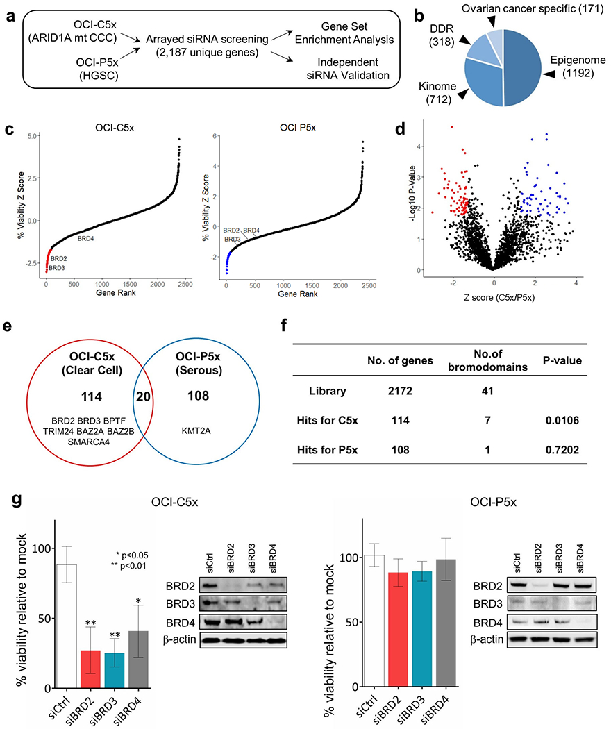
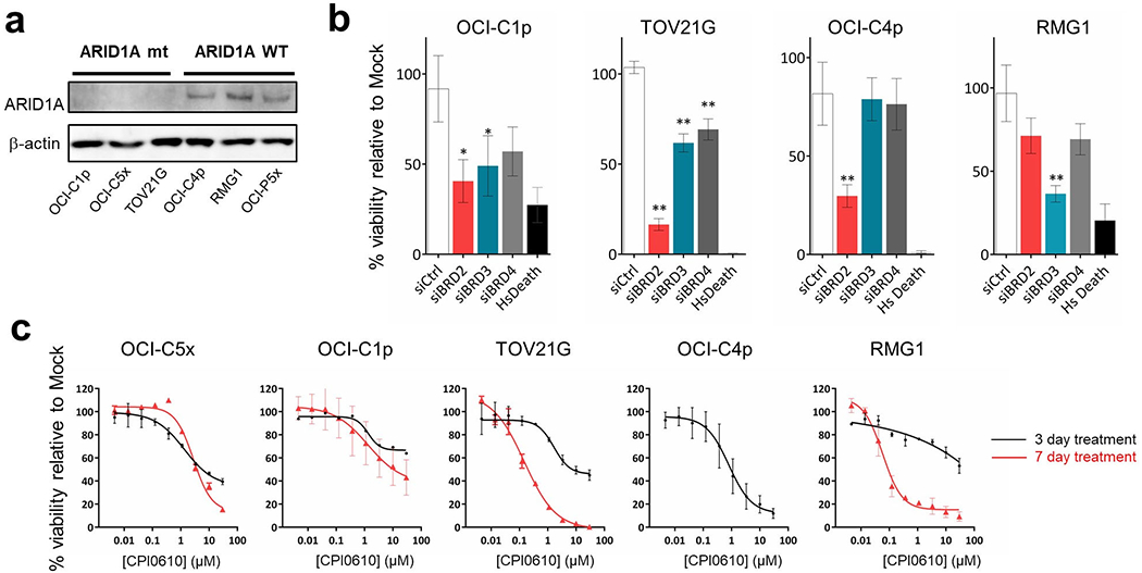
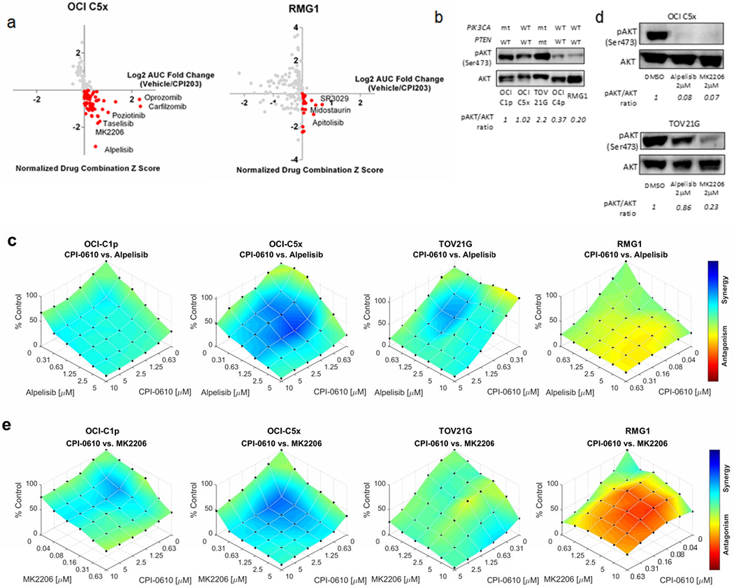
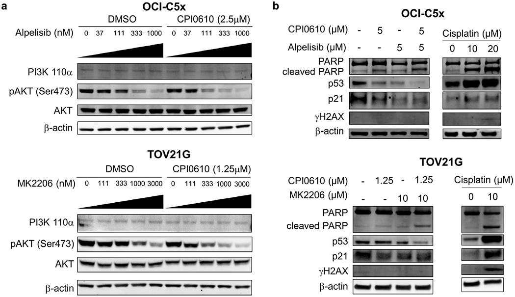
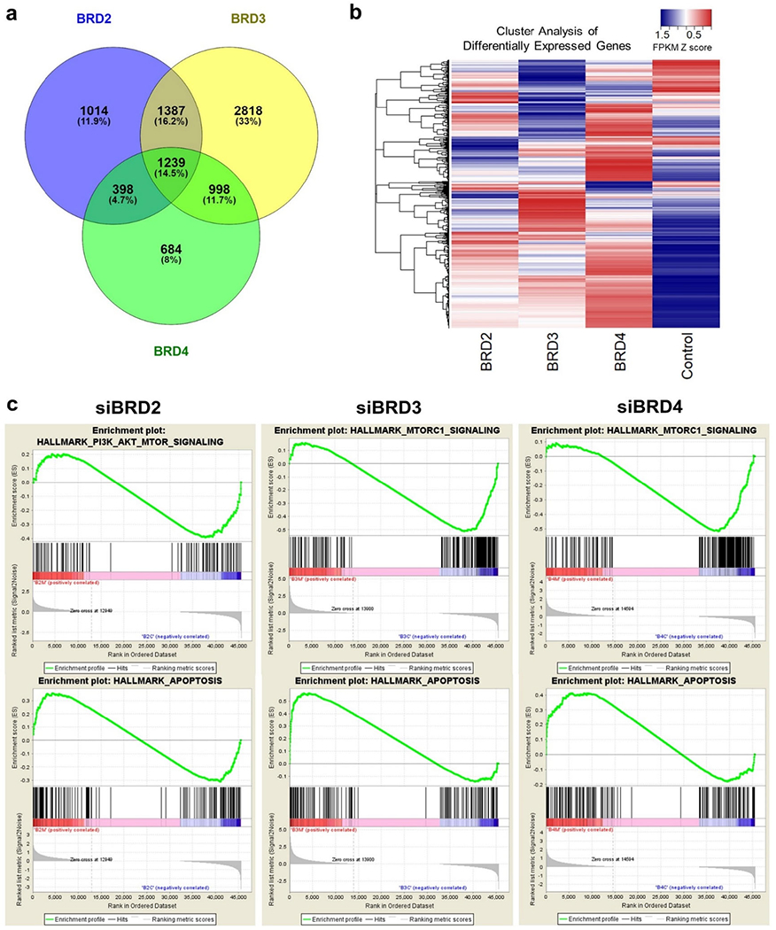
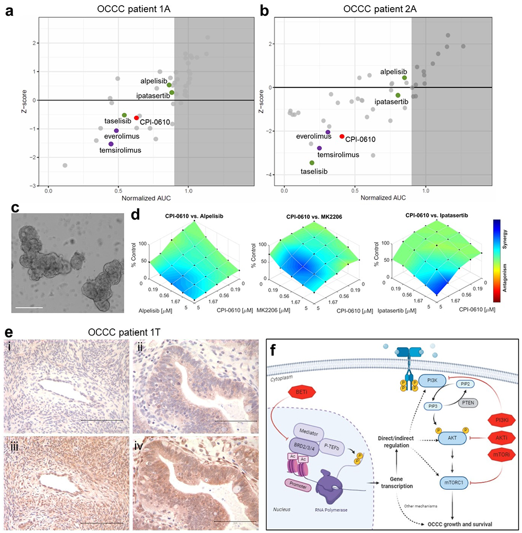
Ovarian clear cell carcinoma (OCCC) is a rare, chemo-resistant subtype of ovarian cancer. To identify novel therapeutic targets and combination therapies for OCCC, we subjected a set of patient-derived ovarian cancer cell lines to arrayed high-throughput siRNA and drug screening. The results indicated OCCC cells are vulnerable to knockdown of epigenetic gene targets such as bromodomain and extra-terminal domain (BET) proteins BRD2 and BRD3. Subsequent RNA interference assays, as well as BET inhibitor treatments, validated these BET proteins as potential therapeutic targets. Because development of resistance to single targeted agents is common, we next performed sensitizer drug screens to identify potential combination therapies with the BET inhibitor CPI0610. Several PI3K or AKT inhibitors were among the top drug combinations identified and subsequent work showed CPI0610 synergized with alpelisib or MK2206 by inducing p53-independent apoptosis. We further verified synergy between CPI0610 and PI3K-AKT pathway inhibitors alpelisib, MK2206, or ipatasertib in tumor organoids obtained directly from patients with OCCC. These findings indicate further preclinical evaluation of BET inhibitors, alone or in combination with PI3K-AKT inhibitors for OCCC, is warranted.


Conflict of interest statement

Conflict of interest: CJK and CG are founders and equity holders in SEngine Precision Medicine. All other authors declare no conflicts of interest.

Figures

Figure 1.
Figure 1.. Arrayed siRNA screens identify bromodomain proteins as targetable vulnerabilities in OCCC.
(a) Scheme of high throughput arrayed siRNA screens. (b) Makeup of custom siRNA library. (c) Z-score based target gene selection. Mean percentage cell viability relative to mock transfection was determined and transformed to Z-score. Genes were sorted by Z-score and plotted. Genes with Z-score less than −1.65 were selected as hits (red or blue) for each OCI cell type. (d) Volcano plot of cell viability comparison between OCI-C5x and OCI-P5x cells. Percentage cell viability was standardized in reference to the mean of each cell and fold change (OCI-C5x/OCI-P5x) was calculated and transformed to Z score. P-values for each gene were also determined by Student t-test. False discovery rate was calculated and FDR>1.29 or <−1.29 was used to select hits specific to OCI-C5x (red) and OCI-P5x (blue), respectively. (e) Venn diagram to summarize the hits from the siRNA screen. Genes encoding bromodomain proteins are listed. (f) Summary table for the number of bromodomain genes in the siRNA library and as hits. P-values were determined by Fisher’s exact test between the library and the hits for OCI C5x or OCI P5x, respectively. (g) Validation assay focusing on BET proteins BRD2, BRD3, and BRD4. Cells were transfected with 10nM of pooled dsiRNAs targeting each BET gene. Cell viability was assessed 72 hours after transfection. Values were the means ± SD of three independent assays. Knockdown efficiency and specificity were confirmed by western blotting.
Figure 2.
Figure 2.. Validation of BET proteins as candidate targets for OCCC.
(a) Western blotting of ARID1A expression. OCCC cells harboring ARID1A mutations (OCI-C1p, OCI-C5x, TOV21G) or wild type ARID1A (OCI-C4p, RMG1, OCI P5x) were examined. The result was validated in three independent assays. (b) Knockdown of BET RNA in additional cell lines. 10nM of pooled dsiRNAs targeting BRD2, BRD3, BRD4 and negative control dsiRNAs were transfected into OCI-C1p, OCI-C4p, TOV21G and RMG1 cells. Cell viability was assessed 72 hours after the transfection. 10nM of AllStars Hs Cell Death Control siRNA (Hs Death) was transfected in parallel as a positive control for successful transfection. Values were the means ± SD of three independent assays. * represents P value <0.05 and ** represents P value < 0.01. (c) Dose-response curves of the BET inhibitor CPI0610 in OCCC cells. Cells were treated with the indicated concentrations of CPI0610 for 72 hours (black) or 7 days (red). Only a dose-response curve after 72 hours was obtained in OCI-C4p as the cells were unstable in long-term culture. Values were the means ± SD of three independent assays.
Figure 3.
Figure 3.. Drug combination assays with BET inhibitors and PI3K-AKT pathway inhibitors.
(a) The result of drug combination screens with CPI203 in OCI-C5x and RMG1 cells. X axis: AUC fold change (Vehicle/CPI203). Y axis: normalized combination Z scores. Inhibitors potentially synergistic with CPI203 are highlighted in red. (b) Western blotting for AKT phosphorylation (phospho-AKT) at serine 473 across multiple OCCC cells. Each blot was quantified by densitometry and relative pAKT/AKT ratio in reference to OCI C1p was determined as shown at the bottom. The reproducibility was verified in three independent assays. (c) Assessment of the synergistic interaction between CPI0610 and alpelisib using the Bliss Independence (BI) model. Cells were treated with CPI0610 and alpelisib at the indicated concentrations for 6 days. The maximum concentration was determined according to the sensitivity of each cell to the inhibitors up to 10μM. (d) Western blotting for phospho-AKT after 24 hours of the treatment with alpelisib or MK2206. Relative pAKT/AKT ratio in reference to DMSO treatment was determined by densitometry. Top; OCI-C5x, bottom; TOV21G. The experiment was repeated three times and the same trend was observed. (e) Assessment of the synergistic interaction between CPI0610 and MK2206 using the Bliss Independence (BI) model. Cells were treated with CPI0610 and MK2206 at the indicated concentrations for 6 days. The maximum concentration of MK2206 was determined as described above.
Figure 4.
Figure 4.. BET inhibition sensitizes OCCC cells to PI3K-AKT inhibitors by promoting p53-independent apoptosis.
(a) OCI-C5x and TOV21G cells were treated with alpelisib or MK2206 at multiple concentrations in combination with CPI0610 or DMSO for 48 hours. Expression of PI3K p110α and phospho-AKT were examined by western blotting. Each experiment was repeated twice independently to validate the reproducibility. (b) Cells were treated with the indicated concentrations of CPI0610 and alpelisib (OCI-C5x) or MK2206 (TOV21G). PARP cleavage, p53 and p21 expression and phosphorylation of histone H2AX at serine 139 was determined by western blotting 48 hours after drug administration. Each experiment was repeated twice independently to validate the reproducibility.
Figure 5.
Figure 5.. mRNA-Seq indicates acute depletion of each BET protein alters the PI3K-AKT signaling pathway.
(a) Venn diagram to summarize the mRNA-Seq results. The number of genes up- or down-regulated by each BET protein knockdown is shown. (b) Overview of the cluster analysis focusing on differentially expressed genes. FPKM was transformed to Z score and cluster analysis was performed. The result was visualized as a heatmap. (c) Results of Gene Set Enrichment Analysis (GSEA). FPKM values in triplicates between each BET protein knockdown and control were compared. Top: The enrichment of PI3K-AKT-MTORC or MTORC1 signaling gene set. Bottom: Gene set enrichment related to apoptosis.
Figure 6.
Figure 6.. Sensitivity to combined BET and PI3K/AKT inhibitors in tumor organoids derived from OCCC patients.
(a, b) The result of high-throughput drug screens in organoids derived from ascites of OCCC patient 1 (1A) and patient 2 (2A). (c) Representative brightfield image of organoids cultured from surgical resection of primary tumor of OCCC patient 1 (1T). Scale bar: 200 μm. (d) Dose-response plots of CPI0610 in combination with alpelisib, MK2206, or ipatasertib demonstrating synergistic effects in organoids derived from tumor sample of OCCC patient 1 (1T). (e) Immunohistochemical staining of tissue sections from OCCC patient 1 (1T): (i) normal ovary tissue stained for p-AKT (Ser473); (ii) tumor tissue stained for p-AKT (Ser473); (iii) normal ovary tissue stained for total AKT; (iv) tumor tissue stained for total AKT. Images are representative of staining across tissue sections and were captured using a 40x lens and 10x objective. Scale bar: 100 μm. (f) Schematic of mechanisms of PI3K and BET inhibitor synergy. Knockdown or inhibition of BET proteins directly or indirectly alters PI3K signaling which synergizes with PIK3CA or AKT inhibition leading to p53-independent apoptosis.

References

    1. Henley SJ, Ward EM, Scott S, Ma J, Anderson RN, Firth AU, et al. Annual report to the nation on the status of cancer, part I: National cancer statistics. Cancer. 2020;126(10):2225–49. - PMC - PubMed
    1. Moore K, Colombo N, Scambia G, Kim BG, Oaknin A, Friedlander M, et al. Maintenance Olaparib in Patients with Newly Diagnosed Advanced Ovarian Cancer. N Engl J Med. 2018;379(26):2495–505. - PubMed
    1. Coleman RL, Oza AM, Lorusso D, Aghajanian C, Oaknin A, Dean A, et al. Rucaparib maintenance treatment for recurrent ovarian carcinoma after response to platinum therapy (ARIEL3): a randomised, double-blind, placebo-controlled, phase 3 trial. Lancet. 2017;390(10106):1949–61. - PMC - PubMed
    1. Moore KN, Secord AA, Geller MA, Miller DS, Cloven N, Fleming GF, et al. Niraparib monotherapy for late-line treatment of ovarian cancer (QUADRA): a multicentre, open-label, single-arm, phase 2 trial. Lancet Oncol 2019;20(5):636–48. - PubMed
    1. Anglesio MS, Carey MS, Kobel M, Mackay H, Huntsman DG, Vancouver Ovarian Clear Cell Symposium S. Clear cell carcinoma of the ovary: a report from the first Ovarian Clear Cell Symposium, June 24th, 2010. Gynecol Oncol. 2011;121(2):407–15. - PubMed

Publication types

MeSH terms