Review
. 2021 Apr;42(4):618-626.
doi: 10.3174/ajnr.A6949. Epub 2021 Jan 28.

Retinoblastoma: What the Neuroradiologist Needs to Know

Affiliations
Review

Retinoblastoma: What the Neuroradiologist Needs to Know

V M Silvera et al. AJNR Am J Neuroradiol. 2021 Apr.

Abstract

Retinoblastoma is the most common primary intraocular tumor of childhood. Accurate diagnosis at an early stage is important to maximize patient survival, globe salvage, and visual acuity. Management of retinoblastoma is individualized based on the presenting clinical and imaging features of the tumor, and a multidisciplinary team is required to optimize patient outcomes. The neuroradiologist is a key member of the retinoblastoma care team and should be familiar with characteristic diagnostic and prognostic imaging features of this disease. Furthermore, with the adoption of intra-arterial chemotherapy as a standard of care option for globe salvage therapy in many centers, the interventional neuroradiologist may play an active role in retinoblastoma treatment. In this review, we discuss the clinical presentation of retinoblastoma, ophthalmic imaging modalities, neuroradiology imaging features, and current treatment options.


Figures

FIG 1.
FIG 1.
A 21-month-old boy with right group B and left group E retinoblastoma. Coronal reformat of 3D FIESTA (A) shows a hypointense calcified mass in the left globe with serous retinal detachment. Two of 3 small retinoblastoma tumors in the right globe are shown (black solid arrows). Axial post-gadolinium T1-weighted image with fat saturation (B) shows enhancing tumor abutting the normally enhancing choroid (white arrow) making focal tumor invasion challenging to assess. Tiny foci of enhancement surrounding the left optic nerve cuff represent normal small vessels (black and white arrow). The left globe was larger than the right consistent with buphthalmos. Gross pathology specimen of the enucleated left eye (C) shows the posteriorly located intraocular neoplasm (arrow). Histopathologic examination showed moderately differentiated retinoblastoma without choroidal invasion and with tumor involving the optic nerve head and lamina cribrosa, but without postlaminar tumor.
FIG 2.
FIG 2.
Group E retinoblastoma managed with IAC and IvitC. A 2-year-old boy presented with leukocoria in the right eye (A). Ultrasonography (B) revealed a large, round, hyperechoic retinal mass with intrinsic calcifications seen as foci of high reflectivity and serous retinal detachment (white arrow). After 3 cycles of IAC and IvitC using melphalan and topotecan, a follow-up sonography image (C) shows regression of tumor to a calcified hyperechoic scar with persistent serous retinal detachment (black arrow).
FIG 3.
FIG 3.
A 21-month-old boy with group E retinoblastoma of the right eye. Axial fat-saturated T2-weighted image (A) shows a large hypointense intraocular tumor (short white arrow) in the right globe with retinal detachment and hemorrhage with a fluid–fluid level (long white arrow). Abnormal thickening of the right optic nerve (arrowhead) is consistent with optic nerve invasion. Sagittal postgadolinium T1-weighted image with fat saturation (B) demonstrates enhancing tumor contacting the lens (short arrow) and tumor invading the optic nerve and extending posteriorly along the optic nerve sheath (long arrow) to the level of the optic canal (open arrow). Histopathologic examination showed poorly differentiated retinoblastoma with choroidal and extensive postlaminar optic nerve invasion.
FIG 4.
FIG 4.
A 2-year-old girl with group E retinoblastoma. Axial fat-saturated T2-weighted image (A) shows a hypointense intraocular tumor centered within the posterior right globe with retinal detachment. Axial trace DWI shows reduced diffusivity of the tumor (B). Axial postgadolinium T1-weighted image with fat saturation (C) shows enhancement of the distal optic nerve in continuity with tumor over 1.2 mm (arrow) and enhancing tumor abutting the enhancing choroid, limiting the assessment for focal choroidal invasion. Histopathologic examination showed poorly differentiated retinoblastoma (grade IV of IV) largely confined to the posterior eye with postlaminar optic nerve invasion and no choroidal invasion.
FIG 5.
FIG 5.
Group D retinoblastoma managed with IAC and IvitC. A 7-year-old boy presented with a large, white inferiorly located orbital tumor shown in the color fundus photograph (A). Ultrasonography revealed a round, hyperechoic retinal mass (B). After 4 cycles of IAC and IvitC using melphalan and topotecan, the tumor regressed to a small, calcified scar seen on fundoscopy (C) and confirmed by ultrasonography (D) (white arrow).
FIG 6.
FIG 6.
Competitive flow to the choroid from the OA and middle meningeal artery (MMA). A lateral angiographic image provides reference for OA supply to the globe (A); the posterior ciliary artery is the vascular supply to the choroid and produces the choroid blush. This is the imaging surrogate for the origin of the central retinal artery. Right ICA injection (B) shows the normal OA origin (black arrow) and expected choroid blush (white arrow). Right ECA injection (C) shows the artery of the superior orbital fissure (Art SOF; black and white arrows), which anastomoses with distal branches of the OA and then fills the OA in a retrograde fashion (black arrows) indicating competitive flow between the MMA and OA for supply to the retina. MR angiogram (D) shows supply to the orbit through the artery of the superior orbital fissure (white arrow), a branch of the MMA (white and black arrow).
FIG 7.
FIG 7.
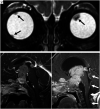
A 16-month-old boy presented with extensive metastatic disseminated quadrilateral retinoblastoma. Coronal T2-weighted image from the initial MR imaging (A) showed hypointense retinal tumors in both globes (black arrows). Sagittal postgadolinium T1-weighted image from the initial MR imaging (B) showed a large enhancing mass occupying the sella, suprasellar cistern, and third ventricle, with separate tumors involving the pineal gland and extending along the planum sphenoidale. Sagittal postgadolinium T1-weighted image from an MR imaging 5 weeks later (C) shows tumor growth, obstructive hydrocephalus, and leptomeningeal dissemination of tumor within the fourth ventricle and along the brain stem and upper cervical cord (white arrows).

Similar articles

Cited by

References

    1. Dimaras H, Kimani K, Dimba EA, et al. . Retinoblastoma. Lancet 2012;379:1436–46 10.1016/S0140-6736(11)61137-9 - DOI - PubMed
    1. Dimaras H, Corson TW, Cobrinik D, et al. . Retinoblastoma. Nat Rev Dis Primers 2015;1:15021 10.1038/nrdp.2015.21 - DOI - PMC - PubMed
    1. Shields JA, Leonard BC, Michelson JB, et al. . B-scan ultrasonography in the diagnosis of atypical retinoblastomas. Can J Ophthalmol 1976;11:42–51 - PubMed
    1. Chawla B, Bhaskaran K, Dada T, et al. . Evaluation of the role of ultrasound biomicroscopy in advanced retinoblastoma: a prospective study on Asian Indian children. Ophthalmic Genet 2020;1–6 10.1080/13816810.2020.1737946 - DOI - PubMed
    1. Kim JW, Ngai LK, Sadda S, et al. . Retcam fluorescein angiography findings in eyes with advanced retinoblastoma. Br J Ophthalmol 2014;98:1666–71 10.1136/bjophthalmol-2014-305180 - DOI - PubMed

LinkOut - more resources