. 2021 Jan 29;371(6528):521-526.
doi: 10.1126/science.aax8819.

MAIT cell activation augments adenovirus vector vaccine immunogenicity

Affiliations

MAIT cell activation augments adenovirus vector vaccine immunogenicity

Nicholas M Provine et al. Science. .

Abstract

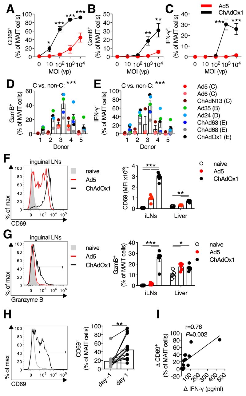
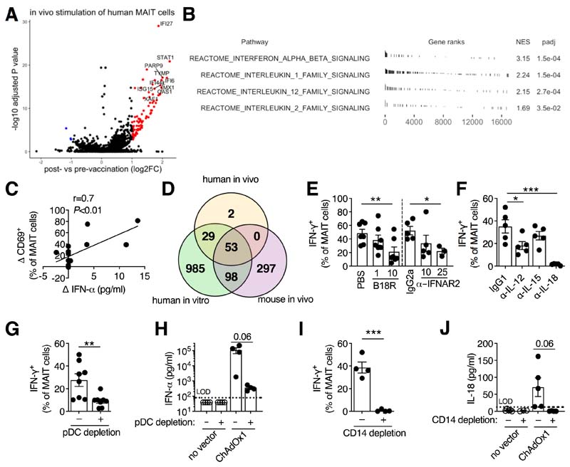
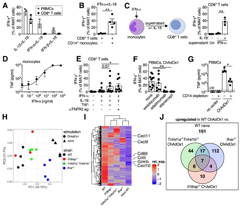
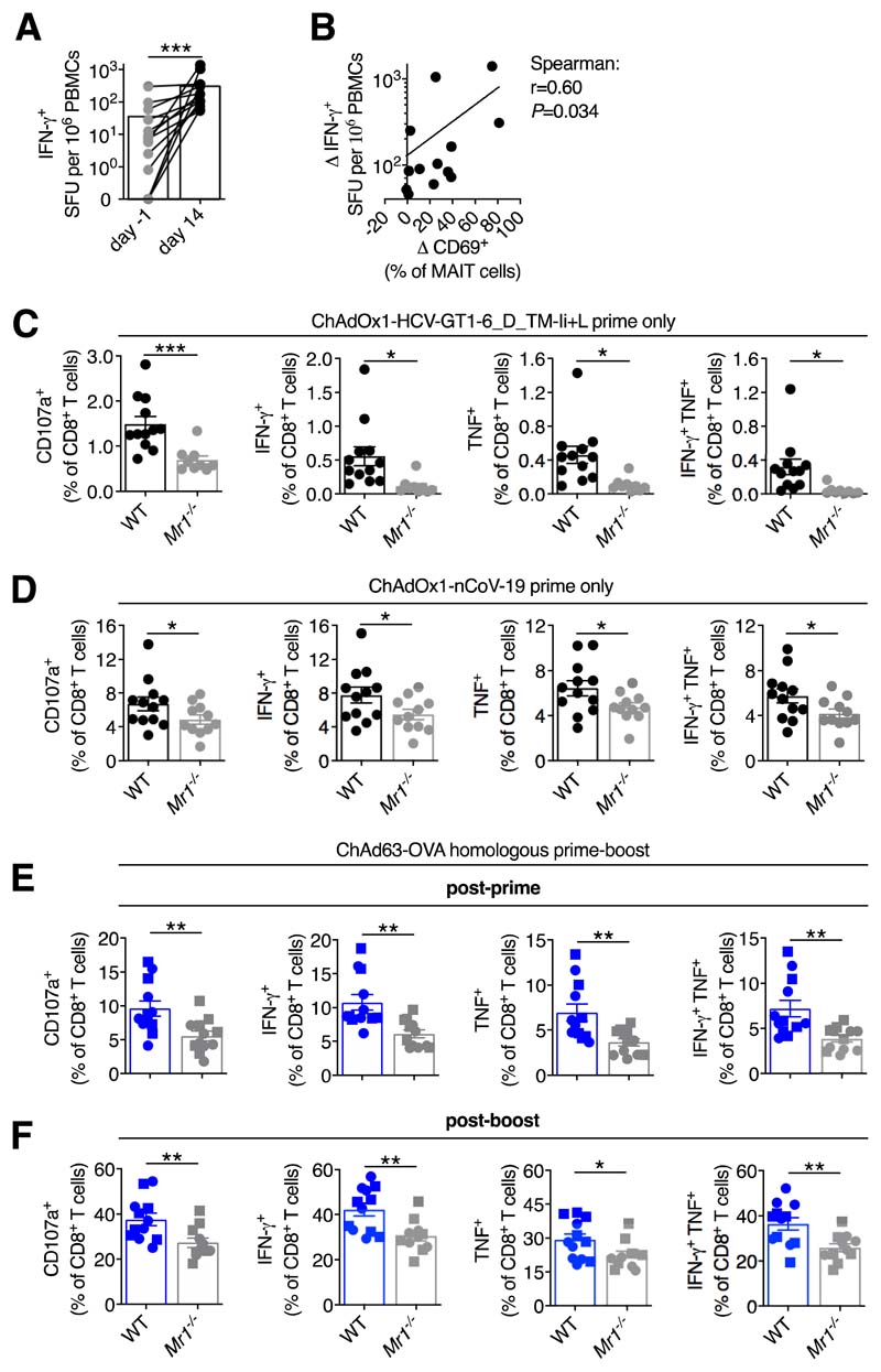
Mucosal-associated invariant T (MAIT) cells are innate sensors of viruses and can augment early immune responses and contribute to protection. We hypothesized that MAIT cells may have inherent adjuvant activity in vaccine platforms that use replication-incompetent adenovirus vectors. In mice and humans, ChAdOx1 (chimpanzee adenovirus Ox1) immunization robustly activated MAIT cells. Activation required plasmacytoid dendritic cell (pDC)-derived interferon (IFN)-α and monocyte-derived interleukin-18. IFN-α-induced, monocyte-derived tumor necrosis factor was also identified as a key secondary signal. All three cytokines were required in vitro and in vivo. Activation of MAIT cells positively correlated with vaccine-induced T cell responses in human volunteers and MAIT cell-deficient mice displayed impaired CD8+ T cell responses to multiple vaccine-encoded antigens. Thus, MAIT cells contribute to the immunogenicity of adenovirus vectors, with implications for vaccine design.


Conflict of interest statement

Competing interests:

C.D., C.S.R., and A.J.P. are named inventors on a patent application in the field of meningococcal vaccines. A.J.P. waives his rights under any patent. P.K. is a named inventor on a patent application in the field of cancer vaccines.

Figures

Figure 1
Figure 1. Activation of human and murine MAIT cells by adenovirus vectors.
(A to C) Human PBMCs (n=9; four experiments) were stimulated with Ad5-GFP or ChAdOx1-GFP (multiplicity of infection (MOI)=0 to 104 vp (viral particles)). MAIT cell CD69 (A), granzyme B (GzmB) (B), and IFN-γ (C) expression was measured after 24 hours. (D and E) Human PBMCs (n=5; two experiments) were stimulated with the indicated vectors (species in parentheses). MAIT cell GzmB (D) or IFN-γ (E) expression were measured after 24 hours. (F and G) C57BL/6J mice (n=6 per group; representative of two experiments) were immunized intramuscularly (i.m.) with 108 IU (infectious units) of Ad5-GFP or ChAdOx1-GFP. Inguinal LN MAIT cell CD69 (F) and GzmB (G) expression was measured after 24 hours. (H and I) Healthy human volunteers (n=14) were immunized with a 5×10¹⁰ vp dose of ChAdOx1 MenB.1. (H) MAIT cell CD69 expression 1 day pre- and 1 day post-immunization. (I) Pearson correlation of change in plasma IFN-γ levels following vaccination with the change in MAIT cell CD69 expression. *, P<0.05; **, P<0.01; ***, P<0.001. Unpaired t test (A to C), two-way ANOVA (D and E), one-way ANOVA with Sidak correction for multiple comparisons (F and G), or Wilcoxon rank-sum test (H). Symbols indicate average response (A to C) or individual mice/volunteers (D to I). Mean ± SEM are shown.
Figure 2
Figure 2. Activation of MAIT cells by adenovirus vectors requires pDC-derived IFN-α and monocyte-derived IL-18.
(A and B) Gene expression analysis of MAIT cells isolated from the PBMCs of human volunteers 1 day pre- and 1 day post-vaccination with ChAdOx1 MenB.1 (n=14). (A) Volcano plot of differentially expressed genes (log2 FC>1, adjusted P<0.05). The top 10 upregulated genes are annotated. (B) Selected cytokine signaling pathways from the Reactome database enriched by Gene Set Enrichment Analysis. (C) Pearson correlation of change in plasma IFN-α level following vaccination with the change in MAIT cell CD69 expression. (D) Overlap of genes upregulated in MAIT cells from ChAdOx1-vaccinated volunteers, from human PBMCs stimulated with ChAdOx1, and from the draining inguinal LNs of ChAdOx1-vaccinated mice. (E and F) Human PBMCs were stimulated with ChAdOx1-GFP and the following inhibitors were used: vaccinia virus-derived type I interferon antagonist B18R (1 or 10 μg/ml; n=7; three experiments) or anti-IFNAR2 antibody (10 or 25 μg/ml; n=5 or 3; two or one experiments, respectively) (E) or anti-IL-12, anti-IL-15, or anti-IL-18 antibodies (10 μg/ml; n=5; two experiments) (F). MAIT cell IFN-γ expression was measured after 24 hours. (G and H) PBMCs were depleted of CD123+ pDCs or left untreated and stimulated with ChAOx1-GFP. MAIT cell IFN-γ expression (n=8; three experiments) (G) or levels of IFN-α in the cell culture supernatant (n=4; one experiment) (H) were measured after 24 hours. (I and J) PBMCs were depleted of CD14+ monocytes or left untreated and stimulated with ChAdOx1-GFP. MAIT cell IFN-γ expression (n=4; two experiments) (I) or IL-18 levels in the supernatant (n=4; three experiments) (J) were measured after 24 hours. *, P<0.05; **, P<0.01; ***, P<0.001. Repeated-measures one-way ANOVA with Dunnett Correction (E and F), or unpaired t test (G to J). Symbols indicate individual donors. Mean ± SEM are shown.
Figure 3
Figure 3. IFN-α acts directly and indirectly through the induction of TNF to activate MAIT cells.
(A) Human PBMCs or purified CD8+ T cells (n=3; one experiment) were stimulated with the indicated cytokines (50 ng/ml). MAIT cell IFN-γ expression was measured after 24 hours. (B) Purified CD8+ T cells ± CD14+ monocytes (n=4; one experiment) were stimulated with IFN-α + IL-18 (50 ng/ml). MAIT cell IFN-γ expression was measured after 24 hours. (C) Purified monocytes (n=3; one experiment) were stimulated with IFN-α (50 ng/ml), or left untreated. After 24 hours, supernatants were transferred ± IL-18 (50 ng/ml) to autologous purified CD8+ T cells. MAIT cell IFN-γ expression was measured after 24 hours. (D) TNF production by IFN-α-treated CD14-purified monocytes was measured after 24 hours (n=3; one experiment). (E) Purified CD8+ T cells (n=10; four experiments) were stimulated with IFN-α and IL-18 ± TNF (50 ng/ml) or anti-TNFR2 agonist antibody (2.5 μg/ml). MAIT cell IFN-γ expression was measured after 24 hours. (F) PBMCs were stimulated with ChAdOx1 and the following inhibitors were added: vedolizumab (anti-α4β7 integrin antibody, n=8; two experiments), adalimumab (anti-TNF antibody, n=11; three experiments), or etanercept (TNFR2-Fc fusion protein, n=8; two experiments) (10 μg/ml). MAIT cell IFN-γ expression was measured after 24 hours. (G) PBMCs ± CD14-depletion were stimulated with ChAdOx1. Concentration of TNF in the supernatant was measured after 24 hours (n=4; one experiment). (H to J) C57BL/6J (n=4), Il18rap−/− (n=3), Tnfrsf1a−/−Tnfrsf1b−/− (n=4), or Ifnar−/− (n=4) mice were immunized intramuscularly with 108 IU of ChAdOx1-GFP. Naive C57BL/6J mice (n=4) were used as a control. After 24 hours, MAIT cells were isolated from the inguinal LNs and sorted for RNA sequencing (one experiment). (H) Principal component analysis. (I) Heatmap of the upregulated differentially expressed genes (log2 FC>1, adjusted P<0.05) between MAIT cells from ChAdOx1-immunized and naive C57BL/6J mice, with all other groups shown for comparison. (J) Overlap of the genes upregulated (log2 FC>1, adjusted P<0.05) in MAIT cells from ChAdOx1-immunized and naïve C57BL/6J mice, and the genes upregulated in MAIT cells from ChAdOx1-immunized C57BL/6J mice as compared to each of the ChAdOx1-immunized knockout strains. *, P<0.05; **, P<0.01. Unpaired t test (B, C and G), repeated-measures one-way ANOVA with Dunnett Correction (E and F). Symbols indicate individual donors. Mean ± SEM are shown.
Figure 4
Figure 4. MAIT cell deficiency impacts on T cell responses following ChAdOx1 or ChAd63 immunization.
(A) Frequency of IFN-γ-producing PBMCs measured by peptide ELISPOT in ChAdOx1 MenB.1 vaccinated volunteers pre-boost (n=14) or day 14 post-boost (n=13). (B) Spearman rank correlation analysis of the change in MAIT cell CD69 expression from pre-boost to day 1 post-boost versus the increase in IFN-γ-producing PBMCs from pre-boost to day 14 post-boost. (C) C57BL/6J (n=12) or Mr1−/− (n=9) mice were immunized intramuscularly (i.m.) with 108 IU of ChAdOx1-HCV-GT1-6_D_TM-Ii+L (two experiments). On day 16, HCV-specific CD107a+, IFN-γ +, TNF+, or IFN-γ +TNF+ CD8+ T cell responses were measured. (D) C57BL/6J (n=12) or Mr1−/− (n=11) mice were immunized i.m. with 108 IU of ChAdOx1-nCoV-19 (two experiments). On day 13, SARS-CoV-2 spike-specific CD107a+, IFN-γ +, TNF+, or IFN-γ +TNF+ CD8+ T cell responses were measured. (E and F) C57BL/6J (n=12) or Mr1−/− (n=12, n=11 post-boost) were primed i.m. with 107 IU of ChAd63-OVA and boosted intravenously on day 28 with 108 IU (squares) or 109 IU (circles) of ChAd63-OVA. SIINFEKL-specific CD107a+, IFN-γ +, TNF+, or IFN-γ +TNF+ CD8+ T cell responses were measured either 3 weeks post-prime (E) or 3 weeks post-boost (F). *, P<0.05; **, P<0.01; ***, P<0.001. Wilcoxon rank-sum test (A), or two-way ANOVA (C to F). Symbols indicate individual volunteers/mice. Mean ± SEM are shown.

Comment in

Similar articles

Cited by

References

    1. Provine NM, Klenerman P. MAIT Cells in Health and Disease. Annu Rev Immunol. 2020;38:203–228. - PubMed
    1. Loh L, et al. Human mucosal-associated invariant T cells contribute to antiviral influenza immunity via IL-18-dependent activation. Proc Natl Acad Sci USA. 2016;113:10133–10138. - PMC - PubMed
    1. van Wilgenburg B, et al. MAIT cells are activated during human viral infections. Nature Communications. 2016;7:11653–11. - PMC - PubMed
    1. van Wilgenburg B, et al. MAIT cells contribute to protection against lethal influenza infection in vivo. Nature Communications. 2018;9:4706. - PMC - PubMed
    1. Vitelli A, et al. Chimpanzee adenoviral vectors as vaccines - challenges to move the technology into the fast lane. Expert Rev Vaccines. 2017;16:1241–1252. - PubMed

Publication types