Review
. 2021 Sep;16(9):1702-1710.
doi: 10.4103/1673-5374.306064.

Astrocytes: a double-edged sword in neurodegenerative diseases

Affiliations
Review

Astrocytes: a double-edged sword in neurodegenerative diseases

Zhi-Bin Ding et al. Neural Regen Res. 2021 Sep.

Abstract

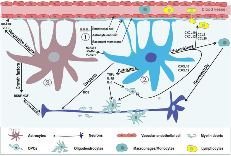
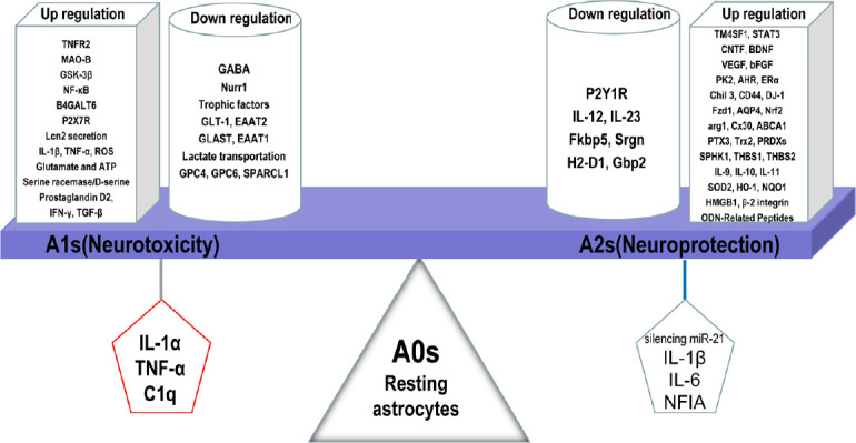
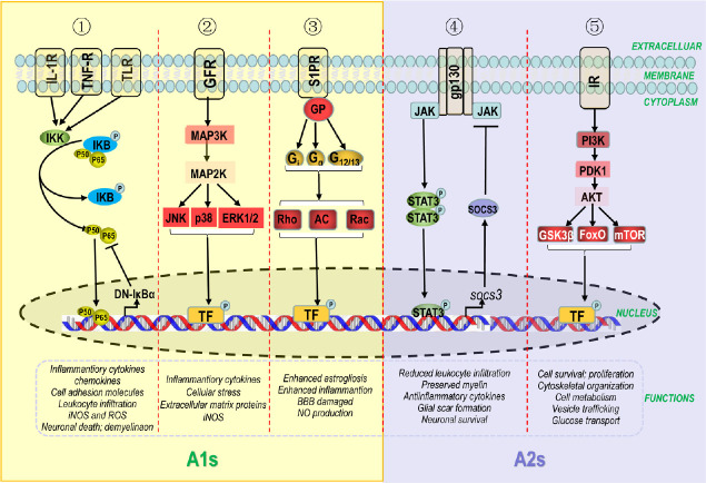
Astrocytes play multifaceted and vital roles in maintaining neurophysiological function of the central nervous system by regulating homeostasis, increasing synaptic plasticity, and sustaining neuroprotective effects. Astrocytes become activated as a result of inflammatory responses during the progression of pathological changes associated with neurodegenerative disorders. Reactive astrocytes (neurotoxic A1 and neuroprotective A2) are triggered during disease progression and pathogenesis due to neuroinflammation and ischemia. However, only a limited body of literature describes morphological and functional changes of astrocytes during the progression of neurodegenerative diseases. The present review investigated the detrimental and beneficial roles of astrocytes in neurodegenerative diseases reported in recent studies, as these cells have promising therapeutic potential and offer new approaches for treatment of neurodegenerative diseases.

Keywords: A1; A2; astrocytes; neurodegenerative diseases; neuroinflammation; neuron; neuroprotection; neurotoxicity; polarization; reactivity.


Conflict of interest statement

None

Figures

Figure 1
Figure 1
Reactive astrocytes in neurodegenerative diseases. Astrocytes can be activated by multiple stimuli, particularly cytokines secreted by different cell types in the brain. In turn, activated astrocytes play a positive or negative role in multiple ways. (1) The blood-brain barrier (BBB) established by the end-feet of astrocytes, capillary endothelial cells, and basement membrane maintains homeostasis of the central nervous system. (2) Reactive astrocytes lose end-feet surrounding capillaries, resulting in damage to the BBB. Meanwhile, reactive astrocytes enhance the release of oxidants, cytokines, chemokines, and cell adhesion molecules, which can directly or indirectly damage oligodendrocyte precursor cells, oligodendrocytes, and neurons. (3) Vasoactive factors secreted by activated astrocytes affect normal endothelial cells by regulating junction-related proteins, which preserves the integrity of the BBB. Moreover, reactive astrocytes can promote remyelination and neuronal regeneration by secreting several growth factors.
Figure 2
Figure 2
Detrimental and beneficial effects of reactive astrocytes induced by various mediators or up/downregulated genes. Resting (A0) astrocytes are stimulated to convert into reactive functional phenotypes (A1 and A2). C1q, IL-1α, and TNF-α secreted by pro-inflammatory microglia transform A0 into A1 astrocytes, which is accompanied by up/downregulation of numerous genes leading to neurodegeneration. In contrast, A2 astrocytes are induced from A0 astrocytes by IL-1β, IL-6, NFIA, and silencing of miR-21, potentially leading to neuroprotective effects by up/downregulation of genes in neurodegenerative disorders. C1q: Component 1 subcomponent q; IL-1α: interleukin-1α; IL-1β: interleukin-1β; IL-6: interleukin-6; NFIA: nuclear factor IA; TNF-α: tumor necrosis factor α.
Figure 3
Figure 3
Intracellular signaling pathways for astrocytic transformation (A1/A2) and roles in neurodegenerative disease. Dysfunctional cells (reactive microglia and neurons) secrete molecules recognized by specific receptors in the astrocytic cytomembrane. These extracellular signals activate intracellular signaling pathways including NF-κB, MAPK, S1PR, JAK/STAT3, and PI3K/AKT. (1) The NF-κB pathway is triggered by inflammatory mediators. Receptor-bound protein kinases activate the IKK complex, resulting in phosphorylation of IκBα, the stable inhibitor of NF-κB. p50 and p65, two subunits of NF-κB, translocate to the nucleus and activate transcription of A1-related genes. DN-IκBα inhibits p50 and p65. (2) The MAPK pathway is partly initiated by cytokines and growth factors, which initiate a cascade amplification of phosphorylation. After triggering, JNK, p38, and ERK1/2 upregulate gene transcription of A1 astrocytes by initiating a large number of transcription factors (TF). (3) The S1PR pathway is activated by extracellular sphingosine-1-phosphate (S1P). S1PR binds to heterotrimeric G-proteins (GPs), such as Gi, Gq and G12/13, which in turn affect downstream proteins such as phospholipase adenylate cyclase (AC), Ras homolog (Rho), Ras-related C3 botulinum toxin substrate (Rac), which translocate to the nucleus to activate expression of A1-related genes. (4) The JAK/STAT3 pathway is triggered by cytokines. Upon cytokine coupling, JAK kinase is initiated and STAT3 is gathered to gp130 (intracellular receptor). Consequently, STAT3 is phosphorylated and translocates to the nucleus, whereby it upregulates gene transcription of A2-related genes. STAT3 also promotes SOCS3 expression, which inhibits the JAK/STAT3 pathway. (5) The PI3K/AKT pathway is activated by binding of insulin to insulin receptor (IR), which activates phosphatidylinositol-4,5-bisphosphate 3-kinase (PI3K). AKT is activated upon its interaction with phosphoinositide dependent protein kinase 1 (PDK1), which is indirectly phosphorylated by PI3K. Subsequently, AKT modulates transcription factors, such as glycogen synthase kinase 3 (GSK3β), forkhead box (FOX), and mechanistic target for rapamycin (mTOR), which are associated with several functions of A2 astrocytes.

References

    1. Acosta C, Anderson HD, Anderson CM. Astrocyte dysfunction in Alzheimer disease. J Neurosci Res. 2017;95:2430–2447. - PubMed
    1. Allaman I, Gavillet M, Belanger M, Laroche T, Viertl D, Lashuel HA, Magistretti PJ. Amyloid-beta aggregates cause alterations of astrocytic metabolic phenotype: Impact on neuronal viability. J Neurosci. 2010;30:3326–3338. - PMC - PubMed
    1. Angelova PR, Abramov AY. Functional role of mitochondrial reactive oxygen species in physiology. Free Radic Biol Med. 2016;100:81–85. - PubMed
    1. Arellano G, Ottum PA, Reyes LI, Burgos PI, Naves R. Stage-specific role of interferon-Gamma in experimental autoimmune encephalomyelitis and multiple sclerosis. Front Immunol. 2015;6:492. - PMC - PubMed
    1. Azam S, Jakaria M, Kim IS, Kim J, Haque ME, Choi DK. Regulation of Toll-Like Receptor (TLR) Signaling pathway by polyphenols in the treatment of age-linked neurodegenerative diseases: focus on TLR4 signaling. Front Immunol. 2019;10:1000. - PMC - PubMed