. 2021 Jan 28;11(1):79.
doi: 10.1038/s41398-021-01205-0.

Altered offspring neurodevelopment in an arginine vasopressin preeclampsia model

Affiliations

Altered offspring neurodevelopment in an arginine vasopressin preeclampsia model

Serena Banu Gumusoglu et al. Transl Psychiatry. .

Abstract

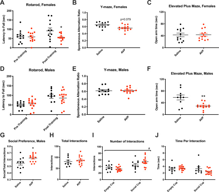
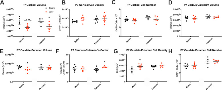
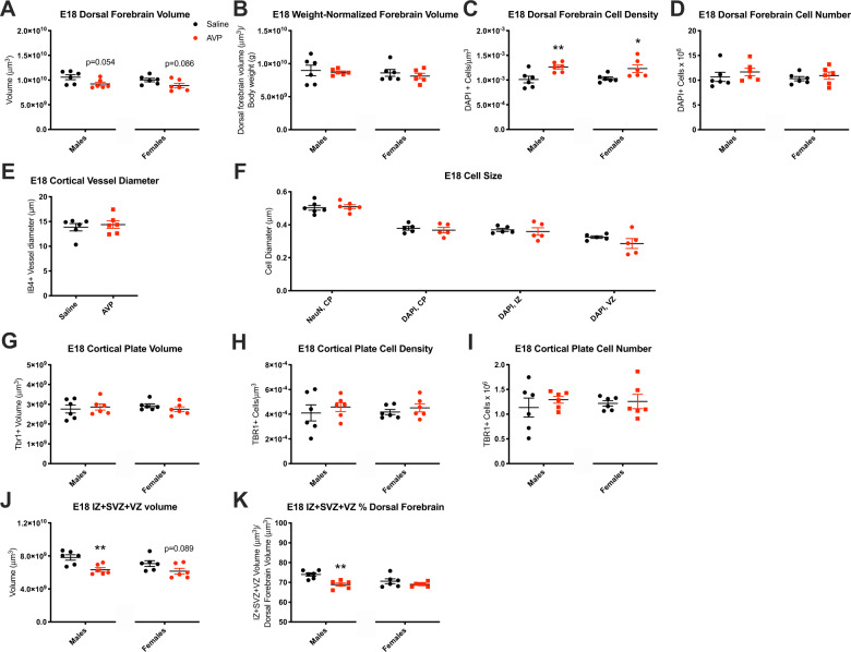
Preeclampsia is a severe gestational hypertensive condition linked to child neuropsychiatric disorders, although underlying mechanisms are unclear. We used a recently developed, clinically relevant animal model of preeclampsia to assess offspring. C57BL/6J mouse dams were chronically infused with arginine vasopressin (AVP) or saline (24 ng/h) throughout pregnancy. Adult offspring were behaviorally tested (Y-maze, open field, rotarod, social approach, and elevated plus maze). Offspring brain was assessed histologically and by RNA sequencing. Preeclampsia-exposed adult males exhibited increased anxiety-like behavior and social approach while adult females exhibited impaired procedural learning. Adult AVP-exposed males had reduced total neocortical volume. Adult AVP-exposed females had increased caudate-putamen volume, increased caudate-putamen cell number, and decreased excitatory synapse density in hippocampal dentate gyrus (DG), CA1, and CA3. At postnatal day 7 (P7), AVP-exposed male and female offspring both had smaller neocortex. At P7, AVP-exposed males also had smaller caudate-putamen volume, while females had increased caudate-putamen volume relative to neocortical size. Similar to P7, E18 AVP-exposed offspring had smaller dorsal forebrain, mainly in reduced intermediate, subventricular, and ventricular zone volume, particularly in males. Decreased volume was not accounted for by cell size or cerebrovascular vessel diameter changes. E18 cortical RNAseq revealed 49 differentially-expressed genes in male AVP-exposed offspring, over-representing cytoplasmic translation processes. In females, 31 genes were differentially-expressed, over-representing collagen-related and epithelial regulation pathways. Gene expression changes in E18 AVP-exposed placenta indicated potential underlying mechanisms. Deficits in behavior and forebrain development in this AVP-based preeclampsia model were distinctly different in males and females, implicating different neurobiological bases.


Conflict of interest statement

M.K.S., D.A.S., and J.L.G. hold patents related to AVP for the prediction and treatment of preeclampsia: US 293 #9,937,182 (April 10, 2018), EU #2,954,324 (July 31, 2019), and PCT/US2018/027152. The authors declare that they have no conflict of interest.

Figures

Fig. 1
Fig. 1. Adult offspring behavior was sex-specifically changed by prenatal maternal AVP.
a AVP-exposed adult females exhibited decreased post-training performance on the rotarod procedural learning task (p = 0.048), while (d) male rotarod learning was unchanged by AVP status. b Trend-significant working memory deficits, as measured by spontaneous alternations on the Y-maze, also occurred in AVP-exposed female (p = 0.079), but not male (e), offspring. c While AVP-exposed female did not exhibit anxiety-like behavior changes, f AVP-exposed male offspring spent significantly decreased time on the open arm of the elevated plus maze (p = 0.0019). g Social preference was significantly (p = 0.018) increased in AVP-exposed male offspring, while (h) total interactions were unchanged. i Offspring from both conditions had significantly more interactions with the social cup than with the empty cup [main effect of cup type by two-way ANOVA p < 0.0001, F1,44 = 19.31], while (j) time spent at each cup did not differ by condition. #p < 0.0001 by two-way ANOVA, *p < 0.05, **p < 0.005 per two-sample t-test, error bars represent SEM. 4 litters per condition, 1–6 offspring per sex per litter.
Fig. 2
Fig. 2. Adult offspring neurobiology was altered by maternal AVP.
a AVP-exposed females had increased caudate–putamen volume (p = 0.037), (b) as well as increased caudate–putamen volume relative to cortex volume (p = 0.021). c Caudate–putamen cell density was unchanged. d The number of cells in the caudate–putamen was trend-increased in AVP-exposed females (p = 0.053). e Caudate–putamen cell numbers were inversely correlated with post-training rotarod performance in adult female offspring (R = −0.6809; p = 0.0014 by exploratory analyses; q = 0.0056 after Benjamini–Hochberg FDR correction). f Excitatory synapse density (PSD95/VGlut1 colocalization) was decreased in the DG in AVP-exposed females (p = 0.046), and (g) trend-decreased in the CA1 (p = 0.053) and decreased in the (h) CA3 (p = 0.030) regions. *p < 0.05 per two-sample t-test, error bars represent SEM. 2–3 litters per condition, 1–3 brains per sex per litter.
Fig. 3
Fig. 3. Juvenile offspring neurobiology was altered by maternal AVP.
a Cortical volume in postnatal day 7 (P7) offspring was trend-smaller in males (p = 0.054) and significantly smaller in AVP-exposed females (p = 0.047). b Cortical cell density and (c) cell number were not changed by AVP at P7, (d) nor was corpus callosum volume, in either sex. e The caudate–putamen was smaller by volume in P7 male AVP-exposed offspring (p = 0.032), while (f) caudate–putamen volume relative to cortical volume was increased among P7 AVP-exposed females (p = 0.043). g The density of cells in the caudate was increased in P7 AVP-exposed males (p = 0.046), resulting in (h) an unchanged number of cells with AVP exposure in either sex. *p < 0.05 per two-sample t-test, error bars represent SEM. 2 litters per condition, 2–3 brains per sex per litter.
Fig. 4
Fig. 4. Embryonic offspring neurobiology was altered by maternal AVP.
a The embryonic day (E) 18 dorsal forebrain was trend-smaller in AVP-condition offspring (males: p = 0.054, females: p = 0.086), which reflected (b) symmetrical growth restriction relative to body weight decrements with AVP exposure. c Dorsal forebrain cell density significantly increased with AVP exposure in male and female offspring [males: p = 0.009, females: p = 0.035] such that (d) the total forebrain cell number was unchanged. These volume differences were not accounted for by (e) changes in cortical vessel diameter or (f) cell size across the cortical plate (CP), intermediate (IZ), or ventricular (VZ) zones. g The TBR1-labeled cortical plate volume, (h) cell density, (i) and cell number were unchanged with AVP. j The volume of the intermediate, subventricular, and ventricular zones (IZ + SVZ + VZ) was significantly smaller in E18 AVP-exposed male offspring and trend-smaller in AVP-exposed female offspring (males: p = 0.005, females: p = 0.089), (k) also reflected by a decreased proportion of dorsal forebrain occupied by IZ + SVZ + VZ in AVP-exposed males (p = 0.001). *p < 0.05, **p < 0.01 per two-sample t-test, error bars represent SEM. 3 litters per condition, 1–3 brains per sex per litter.
Fig. 5
Fig. 5. Transcription indicates E18 cortical sex-specific changes and E18 placental male-specific redox and functional changes with maternal AVP.
a Functional annotation of DE genes with AVP exposure revealed 11 DE biological processes in females and 1 in males with AVP exposure via PANTHERDB. n = 4 per group per sex except n = 3 for female AVP, excluding one outlier for high variance, each from an independent litter. b Embryonic day (E) 18 dorsal forebrain expression of Aquaporin 1 (AQP1) and c Aquaporin 4 (AQP4) were not changed by maternal AVP. d Transcript levels of superoxide dismutase (SOD1) were significantly increased with AVP exposure in E18 placentas of AVP-exposed males (p = 0.005). e Levels of erythroid 2–related factor 2 (NRF2) transcripts were unchanged. f An AVP male-specific decrease occurred in placental nitric oxide synthase 3 (NOS3) transcripts (p = 0.021). g Placental hypoxia-inducible factor 1-alpha (HIF1α) expression increased with AVP exposure in male offspring (p = 0.021). h Insulin-like growth factor 1 (IGF1) expression decreased specifically in males (p = 0.031). *p < 0.05, **p < 0.01 per two-sample t-test, error bars represent SEM.

References

    1. Cleary KL, et al. Use of antihypertensive medications during delivery hospitalizations complicated by preeclampsia. Obstet. Gynecol. 2018;131:441–450. doi: 10.1097/AOG.0000000000002479. - DOI - PMC - PubMed
    1. Podymow T, August P. Update on the use of antihypertensive drugs in pregnancy. Hypertension. 2008;51:960–969. doi: 10.1161/HYPERTENSIONAHA.106.075895. - DOI - PubMed
    1. Gumusoglu SB, Chilukuri ASS, Santillan DA, Santillan MK, Stevens HE. Neurodevelopmental outcomes of prenatal preeclampsia exposure. Trends Neurosci. 2020;43:253–268. doi: 10.1016/j.tins.2020.02.003. - DOI - PMC - PubMed
    1. Dachew BA, Mamun A, Maravilla JC, Alati R. Pre-eclampsia and the risk of autism-spectrum disorder in offspring: meta-analysis. Br. J. Psychiatry. 2018;212:142–147. doi: 10.1192/bjp.2017.27. - DOI - PubMed
    1. Walker CK, et al. Preeclampsia, placental insufficiency, and autism spectrum disorder or developmental delay. JAMA Pediatr. 2015;169:154–162. doi: 10.1001/jamapediatrics.2014.2645. - DOI - PMC - PubMed

Publication types

Substances

LinkOut - more resources