. 2021 May;31(5):542-553.
doi: 10.1038/s41422-021-00468-y. Epub 2021 Jan 28.

Mispair-bound human MutS-MutL complex triggers DNA incisions and activates mismatch repair

Affiliations

Mispair-bound human MutS-MutL complex triggers DNA incisions and activates mismatch repair

Janice Ortega et al. Cell Res. 2021 May.

Abstract

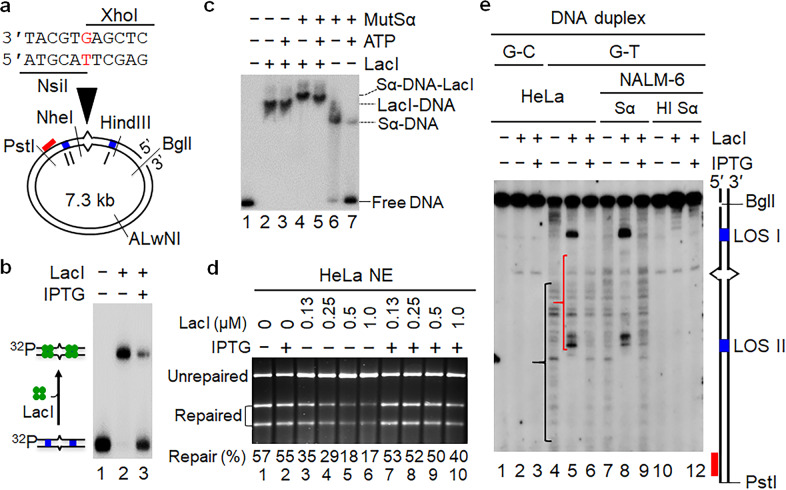
DNA mismatch repair (MMR) relies on MutS and MutL ATPases for mismatch recognition and strand-specific nuclease recruitment to remove mispaired bases in daughter strands. However, whether the MutS-MutL complex coordinates MMR by ATP-dependent sliding on DNA or protein-protein interactions between the mismatch and strand discrimination signal is ambiguous. Using functional MMR assays and systems preventing proteins from sliding, we show that sliding of human MutSα is required not for MMR initiation, but for final mismatch removal. MutSα recruits MutLα to form a mismatch-bound complex, which initiates MMR by nicking the daughter strand 5' to the mismatch. Exonuclease 1 (Exo1) is then recruited to the nick and conducts 5' → 3' excision. ATP-dependent MutSα dissociation from the mismatch is necessary for Exo1 to remove the mispaired base when the excision reaches the mismatch. Therefore, our study has resolved a long-standing puzzle, and provided new insights into the mechanism of MMR initiation and mispair removal.


Conflict of interest statement

The authors declare no competing interests.

Figures

Fig. 1
Fig. 1. Current models of MMR initiation.
a The translocation model suggests that an α-like loop structure forms as a result of the “bidirectional” translocation of MutS homologs when searching for the strand discrimination signal. b The molecular switch model postulates that MSH homologs bind to the mismatch and then slide away from the site to search for the strand discrimination signal in an ATP-dependent manner. c The transactivation model suggests that the MMR initiation complex remains bound to the mismatch and activates downstream nuclease activities at the strand signal via DNA bending/looping.,, d The multi-MLH loading model suggests that mismatch-bound MutS homologs recruit multiple molecules of MutL homologs flanking the mismatch.
Fig. 2
Fig. 2. LacI roadblocks effectively block MutSα sliding.
a The DNA heteroduplex used for in vitro MMR. The circular DNA substrate contains a 5′ nick at the BglI restriction site, a G–T mispair, and two LacI binding sites (blue bars) that are separated by 130 bp. The G–T mismatch was placed in the overlapping recognition sequences of NsiI and XhoI, so that the heteroduplex is resistant to cleavage by both enzymes. The nick-directed MMR removes the mispaired base and subsequent DNA resynthesis restores the sensitivity of the repair product to NsiI, which was used to score for repair. b EMSA assay to determine the specific interaction between LacI and LOS. c EMSA analysis to determine efficient blockage of MutSα sliding by the LacI roadblocks. d In vitro MMR assay showing partial inhibition of MMR in HeLa nuclear extract (NE) by LacI. e Southern blot analysis to determine mismatch-provoked excision intermediates with or without LacI. DNA fragment derived from the circular substrate by a BglI-PstI double digestion contains (from top to bottom) the original strand break, first LacI operon sequence (LOS) I, mismatch, LOS II, and probe annealing site. The red bar represents the 32P-labeled oligonucleotide probe. Sα and HI Sα stand for MutSα and heat-inactivated MutSα, respectively. In all LacI-containing reactions, LacI was preincubated with DNA substrates on ice for 10 min before adding other reaction components.
Fig. 3
Fig. 3. Sliding-deficient MutSα is defective in MMR.
a Amino acid sequences and positions of the Walker A motif in the MSH2 and MSH6 subunits of MutSα. Mutagenesis was focused on methionine (M) and glycine (G) (in blue) in the WT sequence, with the corresponding mutated residues in red. b Structures of Walker A motif of MSH6. Nucleotide-free (upper panel) and ADP-bound (middle panel) MSH6 structures (PDB: 2O8E and 2O8B) are shown in the blue cartoon. The conserved residues and ADP are depicted as sticks and balls and labeled. MG to DA mutations (highlighted in cyan, bottom panel) stabilize the helical conformation, thus making the protein resistant to nucleotide binding. c ATPase activities of WT and mutant MutSα proteins. d ATP-binding activity of WT and mutant MutSα proteins with or without mismatched DNA. Proteins were incubated with [γ-32P]ATP, followed by UV cross-linking and SDS-PAGE. e EMSA analysis to determine ATP-dependent dissociation of WT and mutant MutSα proteins from a 36-bp heteroduplex DNA. f In vitro MMR assay to determine the ability of individual MutSα proteins to restore MMR in MutSα-deficient nuclear extract.
Fig. 4
Fig. 4. Sliding-deficient MutSα triggers mismatch-provoked excision but blocks the excision path at the mismatch site.
a Southern blot analysis to determine mismatch-provoked excision conducted by sliding-deficient MutSα in the reconstituted MMR system with or without the LacI roadblocks, as indicated. The reaction mixtures were incubated at 37 °C for 20 min before being processed for Southern hybridization analysis. b Southern blot analysis to show accumulation of excision intermediates at the mismatch site in the reconstituted MMR system with sliding-deficient MutSα proteins over time. c EMSA analysis to show that the MMR initiation complex contains multiple molecules of MutLα. DNA substrate (0.1 pmol) was a 32P-labeled 100-bp duplex containing a G–T mismatch in the middle. The concentration of MutSα or MutLα used was 2 pmol.
Fig. 5
Fig. 5. MutLα is essential for both 3′- and 5′-directed MMR by nicking the newly synthesized strand 5′ near the mismatch.
a In vitro MMR assay in HeLa nuclear extracts shows that MMR efficiency is inversely proportional to the distance that separates mismatch and strand break. b Principle of mismatch removal assay. c Mismatch removal assay to show that mismatch removal efficiency is the same for all heteroduplexes with various distances between mismatch and strand break. d Mismatch removal assay to show that MutLα endonuclease activity is the determining factor for efficient mismatch removal in heteroduplexes with a long distance between mismatch and strand break. All reactions were incubated at 37 °C for 4 min. e Southern blot analysis shows that MutLα makes multiple incisions (indicated by pink arrows) 5′ to the mismatch, which can effectively remove the mispaired base. f Southern blot analysis shows that LacI roadblocks slightly alter the incision pattern of MutLα, but do not inhibit MutLα endonuclease activity. Green numbers show major incisions 5′ to the mismatch in HeLa nuclear extracts, and their estimated distances (bp) from mismatch are shown in right side of the gel. HL, HeLa nuclear extracts; H6, HCT116 nuclear extracts; EK, a MutLα mutant carrying an E705K substitution in the PMS2 subunit.
Fig. 6
Fig. 6. MutLα interacts with DNA.
ac Sucrose gradient centrifugation was performed to determine the molecular interactions between MutSα, MutLα, and heteroduplex DNA (a), MutSα and DNA (b), MutLα and DNA (c). Reaction mixtures were incubated on ice for 20 min, followed by centrifugation (16 h, 45,000 rpm, at 4 °C), fractionation (from the bottom to the top), electrophoresis, and western blotting.
Fig. 7
Fig. 7. Model of the human MMR process.
Misincorporation can occur in either leading or lagging strand DNA synthesis, and these mispairs can be corrected in a 3′- or 5′-directed manner. Mismatch-bound MutSα recruits MutLα to DNA to form a stable initiation complex, in an ATP-dependent manner. In this ternary complex, mismatch-bound MutSα is flanked by MutLα molecules, and MutLα–MutLα or PCNA–MutLα/MutSα interactions bring mismatch and strand break to proximity, which simplifies the communication between the two sites. MutLα then makes a nick 5′ to the mismatch on the nicked strand. Exo1 is recruited by MutLα to the nick and conducts 5′ → 3′ excision. Once the Exo1-catalyzed excision reaches the mismatch, MutSα or the MutSα–MutLα complex slides away from the mismatch, yielding the right of way to Exo1 for mismatch removal. The excision is terminated by the interactions between MutLα and Exo1. The DNA gap is filled by DNA polymerase δ in concerted reactions with PCNA and RPA, and the nick is ligated by ligase I. This model applies to both 3′ nick-directed (left panel) and 5′ nick-directed (right panel) MMR.

References

    1. Modrich P, Lahue R. Mismatch repair in replication fidelity, genetic recombination, and cancer biology. Annu. Rev. Biochem. 1996;65:101–133. - PubMed
    1. Kolodner RD, Marsischky GT. Eukaryotic DNA mismatch repair. Curr. Opin. Genet. Dev. 1999;9:89–96. - PubMed
    1. Kunkel TA, Erie DA. DNA mismatch repair. Annu. Rev. Biochem. 2005;74:681–710. - PubMed
    1. Li GM. Mechanisms and functions of DNA mismatch repair. Cell Res. 2008;18:85–98. - PubMed
    1. Li F, Ortega J, Gu L, Li GM. Regulation of mismatch repair by histone code and posttranslational modifications in eukaryotic cells. DNA Repair. 2016;38:68–74. - PMC - PubMed

Publication types

LinkOut - more resources