. 2020 Nov 25;30(48):2002931.
doi: 10.1002/adfm.202002931. Epub 2020 Aug 12.

Advanced Materials to Enhance Central Nervous System Tissue Modeling and Cell Therapy

Affiliations

Advanced Materials to Enhance Central Nervous System Tissue Modeling and Cell Therapy

Riya J Muckom et al. Adv Funct Mater. .

Abstract

The progressively deeper understanding of mechanisms underlying stem cell fate decisions has enabled parallel advances in basic biology-such as the generation of organoid models that can further one's basic understanding of human development and disease-and in clinical translation-including stem cell based therapies to treat human disease. Both of these applications rely on tight control of the stem cell microenvironment to properly modulate cell fate, and materials that can be engineered to interface with cells in a controlled and tunable manner have therefore emerged as valuable tools for guiding stem cell growth and differentiation. With a focus on the central nervous system (CNS), a broad range of material solutions that have been engineered to overcome various hurdles in constructing advanced organoid models and developing effective stem cell therapeutics is reviewed. Finally, regulatory aspects of combined material-cell approaches for CNS therapies are considered.

Keywords: central nervous system; materials; organoids; stem cell; therapeutics.


Conflict of interest statement

Conflict of Interest D.V.S. and R.J.M. are inventors on patents related to stem cell manufacturing, and D.V.S., R.J.M., R.G.S., and H.J.J. are co-founders of a company that works on stem cell manufacturing.

Figures

Figure 1.
Figure 1.
Applications of advanced materials in CNS study and repair.
Figure 2.
Figure 2.
Comparison of features for different models of human CNS development.
Figure 3.
Figure 3.
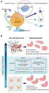
A) Extrinsic regulation of stem cell fate. B) The use of material scaffolds to guide chemical and physical extrinsic regulation across space and time for improved generation of organoids.
Figure 4.
Figure 4.
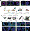
Fully defined material scaffold approaches toward guided organization of organoids. A) Dorso-ventral patterning (SHH and Pax6 expression) in pluripotent stem cell derived neural tube models can be induced by defined dosages of retinoic acid using fully defined laminin and PEG scaffolds in comparison to ill-defined Matrigel scaffold; Reproduced under the terms and conditions of a Creative Commons BY-NC-ND license. Copyright 2014 The Authors, published by Elsevier. B) 60 d protocol (panel a) using a PLGA microfilament scaffold enables physical and spatial patterning for guided cerebral organoid growth (panels b and c) and modified spatial cellular patterning and marker expression compared to an unguided spheroid patterning approach (panels d and e). Adapted with permission.[55] Copyright 2017, Springer Nature.
Figure 5.
Figure 5.
Microscale and macroscale considerations for engineering materials to enhance stem cell manufacturing.
Figure 6.
Figure 6.
Materials can be engineered to overcome problems peri- and post-implantation of neural cells into the CNS.
Figure 7.
Figure 7.
Engineered materials designed to scale up stem cell manufacturing and differentiation to generate desired cell types to target neurological diseases. A) iPSCs are able to expand in 3D PNIPAAm-PEG hydrogels and B) differentiate into ventral midbrain neurons. Adapted under the terms and conditions of a CC BY license.[80] Copyright 2017, Springer Nature Limited. C) H9-hESCs expand in alginate tubes and D,E) retain their pluripotency after long-term culture in these tubes. Adapted under the terms of a CC BY license.[77] Copyright 2018, IOP Publishing.
Figure 8.
Figure 8.
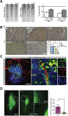
Engineered materials applied toward solving several problems in neural cell transplantation in the central nervous system. A) Cells suspended in thermo-reversible DCH-T at different time points after syringe loading to visually monitor sedimentation in the syringe compared to control cells suspended in cell media. Cell survival is quantified post-injection when suspended in DCH-T and cell media; Adapted with permission from ref. [118]. Copyright 2015 American Chemical Society. B) Host microglial immune response (Iba1+ cells at graft site) and quantification comparing implantation of hMgSCs in PBS and R-GSIK; Adapted with permission.[138] Copyright 2019, Elsevier. C) Vascularization within lesion site measured by CD31+ expression (red) within an iPSC-NPC graft (green) implanted with hyaluronic acid (blue); Reproduced with permission.[141] Copyright 2016, Elsevier. D) HNCAM+ cells (green) implanted in a hyaluronic acid gel functionalized with growth factors (panels g and i) displayed increased graft area and cell innervation post-implantation into the striatum of a rat model of Parkinson’s disease compared to cells implanted in PBS (panels h and j) and corresponding quantification of graft area (panel k); Adapted with permission.[109] Copyright 2018, Wiley-VCH.
Figure 9.
Figure 9.
Roadmap of components to consider when designing a cell therapy with material delivery vehicle.

Similar articles

Cited by

References

    1. Schafer DP, Stevens B, Cold Spring Harbor Perspect. Biol. 2015, 7, a020545. - PMC - PubMed
    1. Noctor SC, Martínez-Cerdeño V, Ivic L, Kriegstein AR, Nat. Neurosci. 2004, 7, 136. - PubMed
    1. Nestler EJ, Hyman SE, Nat. Neurosci. 2010, 13, 1161. - PMC - PubMed
    1. Xiong Y, Mahmood A, Chopp M, Nat. Rev. Neurosci. 2013, 14, 128. - PMC - PubMed
    1. Takahashi K, Tanabe K, Ohnuki M, Narita M, Ichisaka T, Tomoda K, Yamanaka S, Cell 2007, 131, 861. - PubMed