Case Reports
. 2021 Jan 17;16(3):736-741.
doi: 10.1016/j.radcr.2021.01.014. eCollection 2021 Mar.

Granulomatosis with polyangiitis presenting as a solitary renal mass: A case report with imaging and literature review

Affiliations
Case Reports

Granulomatosis with polyangiitis presenting as a solitary renal mass: A case report with imaging and literature review

Takahiro Yamamoto et al. Radiol Case Rep. .

Erratum in

Abstract

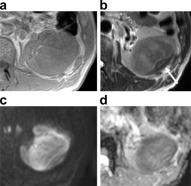
Granulomatosis with polyangiitis (GPA) manifests as necrotizing granulomatous inflammatory masses in the nasal cavity, paranasal sinuses, and lungs. However, a mass in the kidney is extremely rare. We herein report a case of GPA that presented as a solitary mass in the left kidney. The patient was a man in his 60s. A 2.5-cm solitary mass was incidentally discovered in the left kidney at another hospital and was followed-up. Eight months later, the mass had enlarged, and the patient visited our hospital for further examination and treatment. The mass was hypovascular, with unclear margins on contrast-enhanced computed tomography (CT). The signal of the mass was nonuniform and iso- to slightly hypo-intense on T2-weighted and diffusion-weighted magnetic resonance imaging (MRI). Enlarged para-aortic lymph nodes were also detected on the CT and MRI. Based on imaging, malignant tumors were suspected, and nephrectomy was performed. The pathological diagnosis was GPA. We performed a literature review of this rare renal manifestation and present a summary of reported imaging findings. If a hypovascular renal mass with an unclear margin can be found in those with GPA, unnecessary operations may be avoided by actively promoting renal biopsy.

Keywords: CT; Granulomatosis with polyangiitis; Hypovascular renal mass; MRI.


Figures

Fig 1
Fig. 1
Dynamic contrast-enhanced computed tomography. (a) Unenhanced, (b) corticomedullary phase, and (c) nephrogenic phase. There is a 7 cm × 5.5 cm × 6 cm mass in the center of the left kidney (arrow). The contrast effect is weak and incremental: (a) 44 HU, (b) 62 HU, and (c) 72 HU. The border between the mass and renal parenchyma is unclear.
Fig 2
Fig. 2
(a) Contrast-enhanced computed tomography (CT) image taken at another hospital 8 months ago. (b and c) Contrast-enhanced CT images taken at our hospital. The tumor grew from 2.5 cm to 7 cm in length, in 8 months. Poor-contrast area inside of the mass (b), and the ipsilateral para-aortic lymph node is enlarged (c, arrow).
Fig 3
Fig. 3
Magnetic resonance imaging (3.0 T). (a) T1-weighted image shows iso-intense mass relative to the kidney. (b) T2-weighted image shows nonuniform iso- to hypo-intense mass relative to the kidney. There is no pseudo-capsule around the mass. A convex lens-shaped strong low-signal area is found in renal subcapsule (arrow). (c) Diffusion-weighted image (b = 1,000 s/mm2) presents nonuniform and iso- to slightly high-intensity. (d) Apparent diffusion coefficient (ADC) map presents nonuniform and iso- to slightly hypo- intensity, with ADC values ranging from 0.84 × 10−3 to 1.45 × 10−3 mm2/sec.
Fig 4
Fig. 4
Resected specimen (a) and pathological specimens (b-d). (a) Macroscopically, a 9 cm × 7.5 cm yellowish white mass is found in the center of the kidney. Resected specimen shows thickening of the renal capsule in contact with the GPAs and marked thickening of part of the capsule (arrow). Hematoxylin and eosin staining. (b) 20 × magnification, (c) 40 × magnification, and (d) 100 × magnification. Histologically, the lesion shows granuloma with extensive fibrosis, necrosis, and aggregation of neutrophils (b). Granuloma was noted in the renal capsule with markedly thickening fibrosis (a, arrow), (c). Vasculitis with fibrinoid degeneration was confirmed in the small arteries around the ureter (d).

References

    1. Greco A, Marinelli C, Fusconi M, Macri GF, Gallo A, De Virgilo A. Clinic manifestations in granulomatosis with polyangiitis. Int J Immunopathol Pharmacol. 2016;29(2):151–159. doi: 10.1177/0394632015617063. - DOI - PMC - PubMed
    1. Bacon PA. The spectrum of Wegener's granulomatosis and disease replace. N Engl J Med. 2005;325:330–332. doi: 10.1056/NEJMp048338. - DOI - PubMed
    1. Jennette JC, Falk RJ, Bacon PA, Basu N, Cid MC, Ferrario F. 2012 Revised International Capel Hill consensus conference nomenclature of vasculitides. Arthritis Rheum. 2013;65:1–11. doi: 10.1002/art.37715. - DOI - PubMed
    1. Watts RA, Scott DG. ANCA vasculitis: to lump or split? Why we should study MPA and GPA separately. Rheumatol Oxf Engl. 2012;51:2115–2117. doi: 10.1093/rheumatology/kes230. - DOI - PubMed
    1. Fujimoto S, Watts RA, Kobayashi S, Suzuki K, Jayne DR, Scott DG. Comparison of the epidemiology of anti-neutrophil cytoplasmic antibody-associated vasculitis between Japan and U.K. Rheumatol Oxf Engl. 2011;50:1916–1920. doi: 10.1093/rheumatology/ker205. - DOI - PubMed

Publication types

LinkOut - more resources