

Nanofibrous ε-polycaprolactone scaffolds containing Ag-doped magnetite nanoparticles: Physicochemical characterization and biological testing for wound dressing applications in vitro and in vivo
- PMID: 33511308
- PMCID: PMC7809176
- DOI: 10.1016/j.bioactmat.2020.12.026
Nanofibrous ε-polycaprolactone scaffolds containing Ag-doped magnetite nanoparticles: Physicochemical characterization and biological testing for wound dressing applications in vitro and in vivo
Erratum in
-
Erratum regarding missing ethics approval and consent to participate statements in previously published articles.Bioact Mater. 2024 Jun 14;40:275-279. doi: 10.1016/j.bioactmat.2024.06.006. eCollection 2024 Oct. Bioact Mater. 2024. PMID: 38973994 Free PMC article.
Abstract
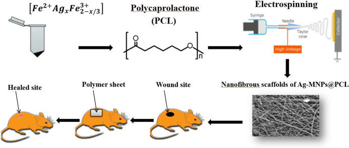
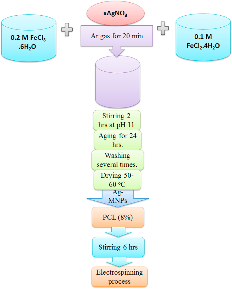
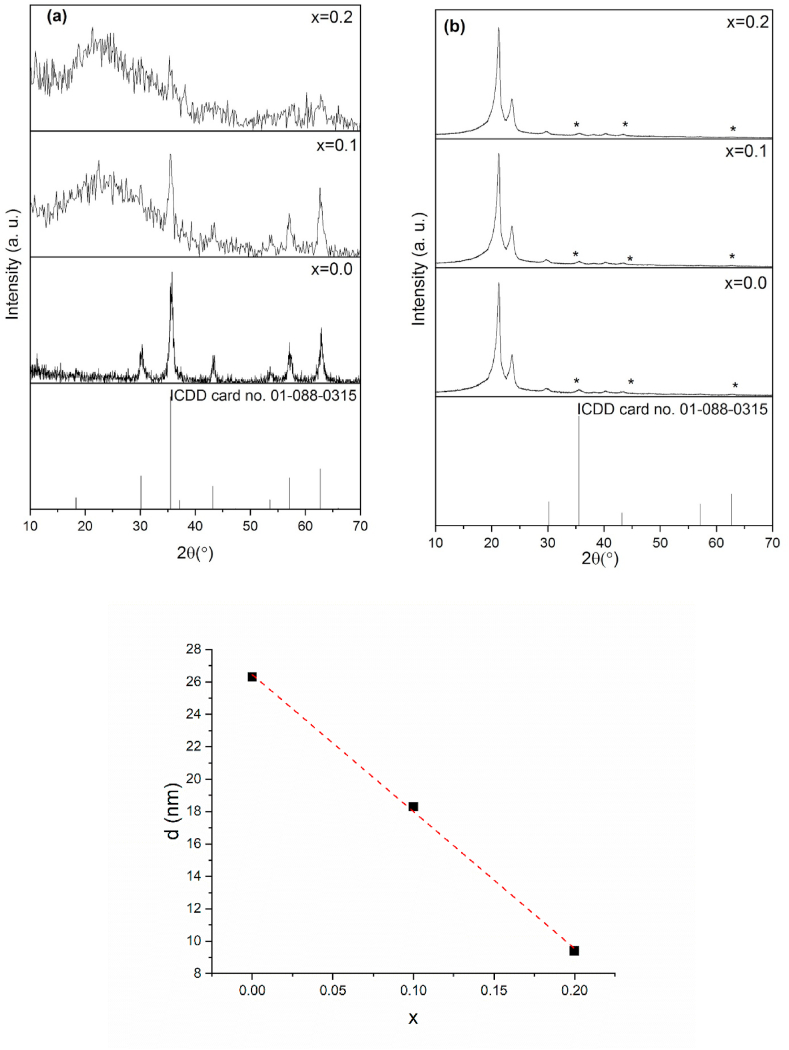
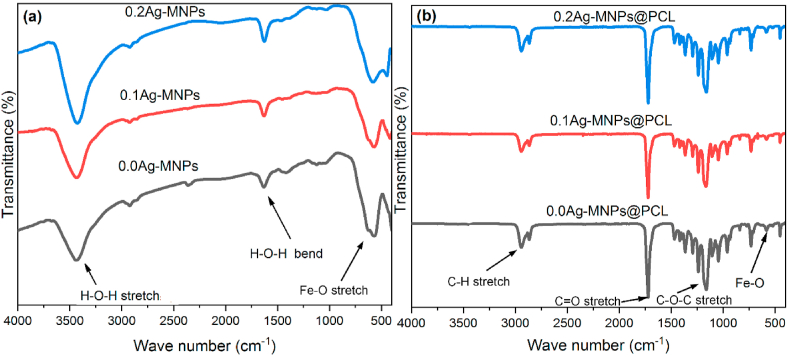
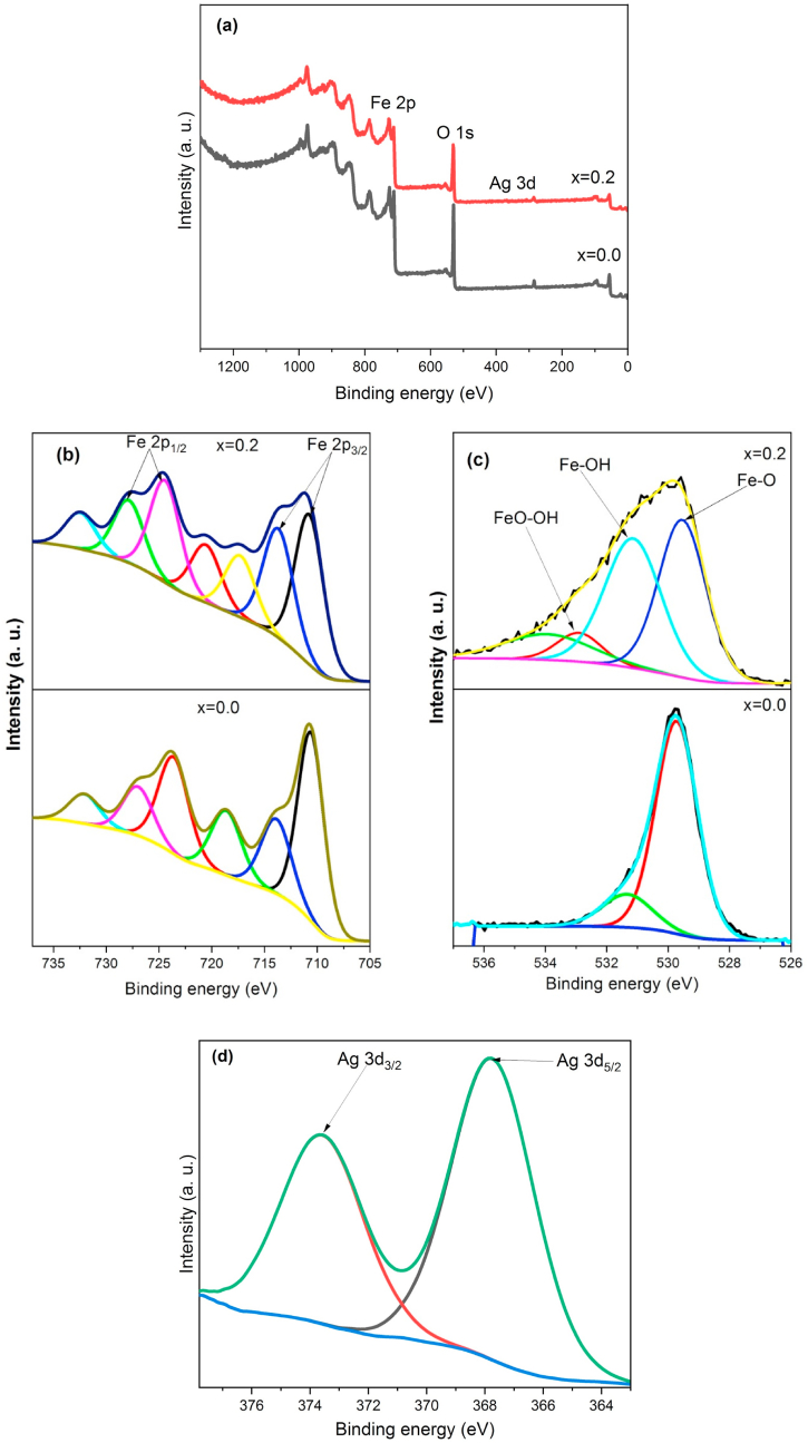
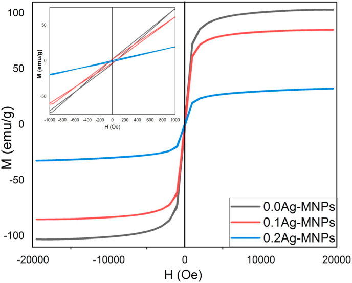
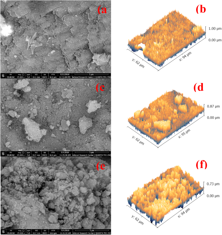
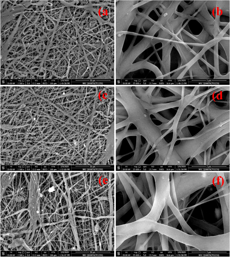
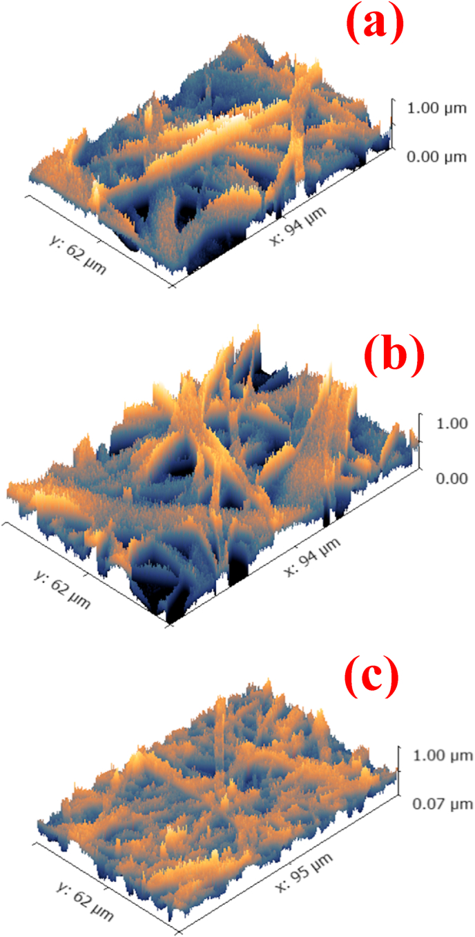
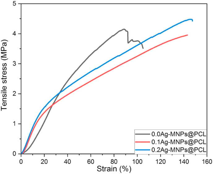
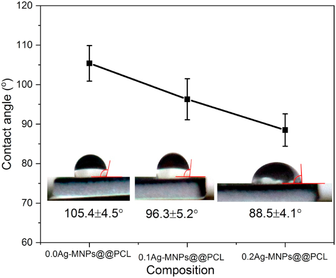
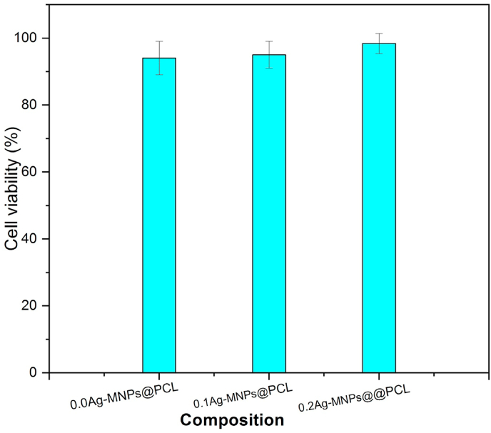
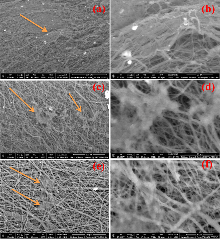
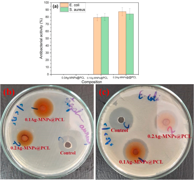
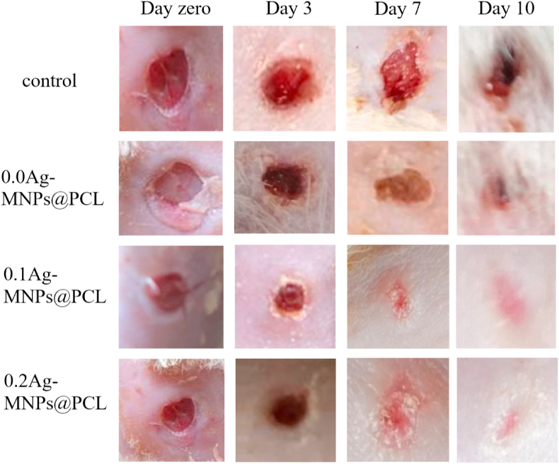
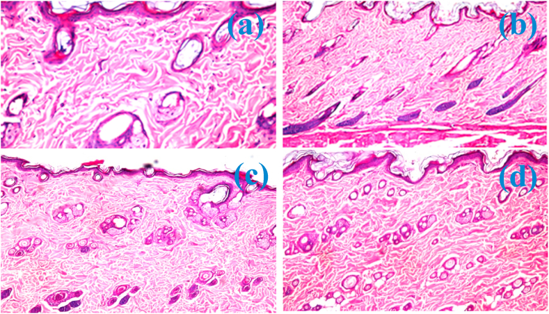
Skin wounds can lead to numerous complications with dangerous health consequences. In this work, magnetite nanoparticles were doped with different concentrations of antimicrobial silver (Ag) ions and incorporated into the electrospun nanofibrous ε-polycaprolactone (PCL) scaffolds. Nanoparticles and scaffolds with various Ag contents were characterized using a range of physicochemical techniques. Ag entered magnetite as cations and preferentially positioned at tetrahedral sites, introducing lattice distortions and topographic irregularities. Amorphization of the structure due to accommodation of Ag expanded the lattice in the bulk and contracted it on the surface, where broadened distribution of Fe-O coordinations was detected. Promoting spin canting and diminishing the double exchange interaction through altered distribution of ferric and ferrous ions, Ag softened the magnetism of magnetite. By making the nanoparticle structure more defective, Ag modified the interface with the polymer and promoted the protrusion of the nanoparticles from the surface of the polymeric nanofibers, thus increasing their roughness and hydrophilicity, with positive repercussions on cell adhesion and growth. Both the viability of human melanocytes and the antibacterial activity against E. coli and S. aureus increased with the concentration of Ag in the magnetite phase of the scaffolds. Skin wound healing rate in rats also increased in direct proportion with the concentration of Ag in the magnetite phase, and no abnormalities in the dermal and epidermal tissues were visible on day 10 in the treatment group. These results imply an excellent potential of these composite nanofibrous scaffolds for use as wound dressings and in other reconstructive skin therapies.
Keywords: Magnetic nanoparticles; Nanofiber; Skin; Tissue engineering; Wound healing.
© 2021 The Authors. Production and hosting by Elsevier B.V. on behalf of KeAi Communications Co., Ltd.
Conflict of interest statement
The authors declare that they have no known competing financial interests or personal relationships that could have appeared to influence the work reported in this paper.
Figures
References
-
- Rivero G., Meuter M., Pepe A., Guevara M.G., Boccaccini A.R., Abraham G.A. Nanofibrous membranes as smart wound dressings that release antibiotics when an injury is infected. Colloid. Surface. Physicochem. Eng. Aspect. 2020;587
-
- Homaeigohar S., Boccaccini A.R. Antibacterial biohybrid nanofibers for wound dressings. Acta Biomater. 2020;107:25–49. - PubMed
-
- Rahimi M., Noruzi E.B., Sheykhsaran E., Ebadi B., Kariminezhad Z., Molaparast M., et al. Carbohydrate polymer-based silver nanocomposites: recent progress in the antimicrobial wound dressings. Carbohydr. Polym. 2020;231 - PubMed
-
- Liu F., Li X., Wang L., Yan X., Ma D., Liu Z., et al. Sesamol incorporated cellulose acetate-zein composite nanofiber membrane: an efficient strategy to accelerate diabetic wound healing. Int. J. Biol. Macromol. 2020;149:627–638. - PubMed
-
- Esmaeili E., Eslami-Arshaghi T., Hosseinzadeh S., Elahirad E., Jamalpoor Z., Hatamie S., et al. The biomedical potential of cellulose acetate/polyurethane nanofibrous mats containing reduced graphene oxide/silver nanocomposites and curcumin: antimicrobial performance and cutaneous wound healing. Int. J. Biol. Macromol. 2020;152:418–427. - PubMed
LinkOut - more resources
Full Text Sources
Other Literature Sources
