. 2021 Jan 11;6(7):2089-2104.
doi: 10.1016/j.bioactmat.2020.12.014. eCollection 2021 Jul.

Cellulose fibers-reinforced self-expanding porous composite with multiple hemostatic efficacy and shape adaptability for uncontrollable massive hemorrhage treatment

Affiliations

Cellulose fibers-reinforced self-expanding porous composite with multiple hemostatic efficacy and shape adaptability for uncontrollable massive hemorrhage treatment

Yansen Wang et al. Bioact Mater. .

Erratum in

Abstract

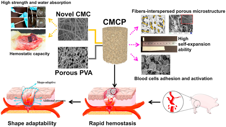
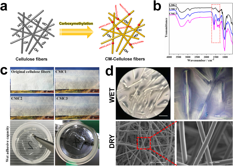
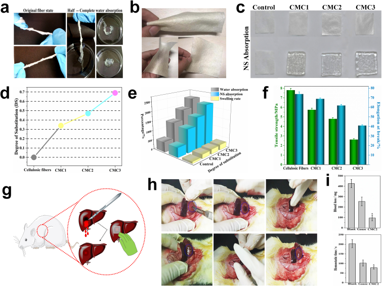
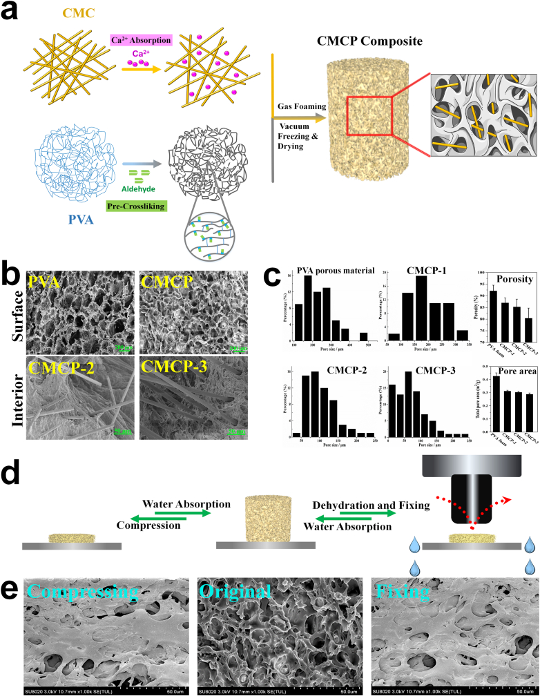
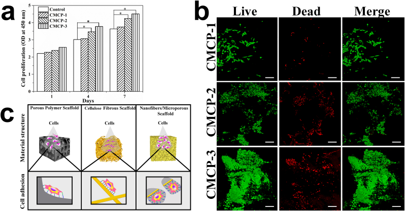
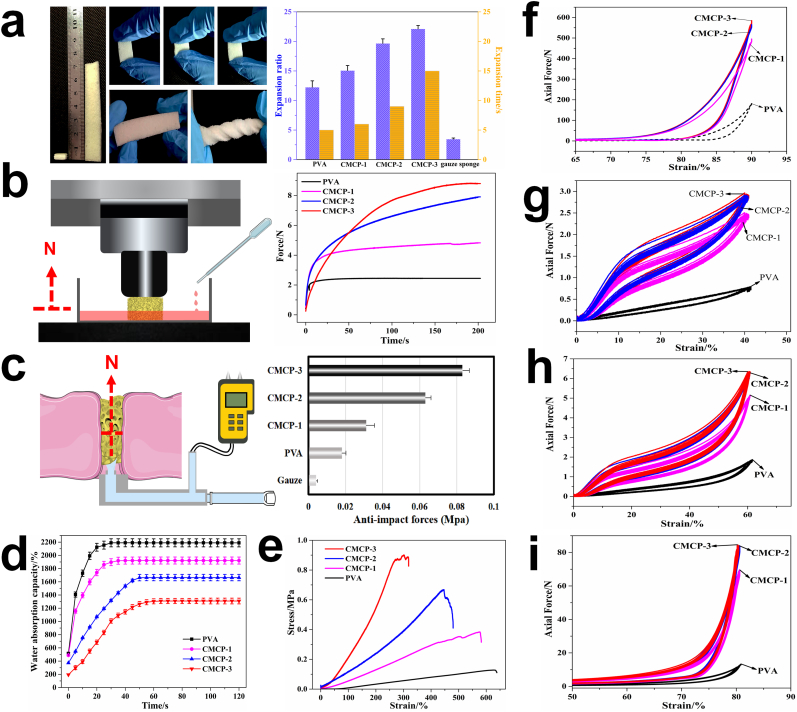
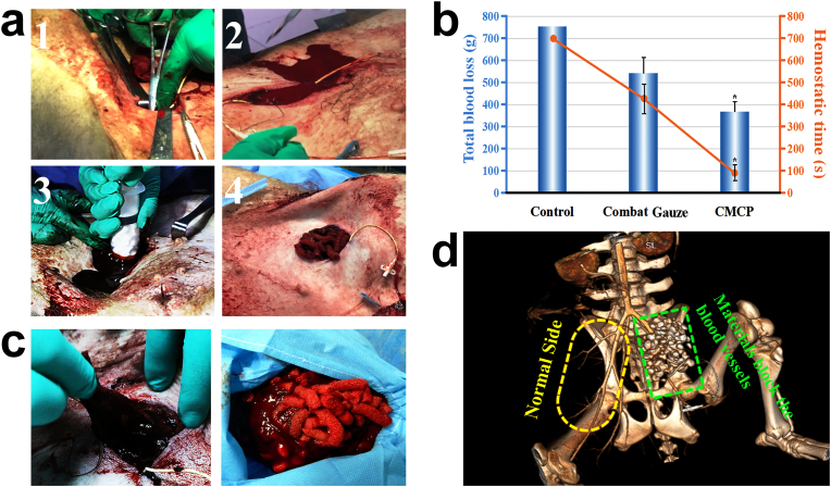
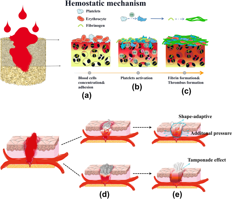
Uncontrollable hemorrhage leads to high mortality and thus effective bleeding control becomes increasingly important in the military field and civilian trauma arena. However, current hemostats not only present limitation when treating major bleeding, but also have various side effects. Here we report a self-expanding porous composites (CMCP) based on novel carboxymethyl cellulose (CMC) fibers and acetalized polyvinyl alcohol (PVA) for lethal hemorrhage control. The CMC fibers with uniform fibrous structure, high liquid absorption and procoagulant ability, are evenly interspersed inside the composite matrix. The obtained composites possess unique fiber-porous network, excellent absorption capacity, fast liquid-triggered self-expanding ability and robust fatigue resistance, and their physicochemical performance can be fine-tuned through varying the CMC content. In vitro tests show that the porous composite exhibits strong blood clotting ability, high adhesion to blood cells and protein, and the ability to activate platelet and the coagulation system. In vivo hemostatic evaluation further confirms that the CMCP presents high hemostatic efficacy and multiple hemostatic effects in swine femoral artery major hemorrhage model. Additionally, the CMCP will not fall off from the injury site, and is also easy to surgically remove from the wound cavity after the hemostasis. Importantly, results of CT tomography and 3D reconstruction indicate that CMCP can achieve shape adaptation to the surrounding tissues and the wound cavities with different depths and shapes, to accelerate hemostasis while protecting wound tissue and preventing infection.

Keywords: Cellulose fibers; Hemostasis; Porous materials; Self-expanding ability; Shape-adaptive.


Figures

Image 1
Graphical abstract
Fig. 1
Fig. 1
a. Schematic diagram of the fabrication of CMC fibers. b. FTIR spectra of different CMC samples. c. Macroscopic observation of different cellulose samples (5 cm × 12 cm) and the CMC samples (2 cm × 2 cm) in wet state. d. The microstructure of the CMC in dry and wet state.
Fig. 2
Fig. 2
a. The original morphology of the CMC and the change of the morphology during water absorption. b. The CMC fibers can be spun into fiber cloth (10 cm × 10 cm) with excellent flexibility. c. Macroscopic images of cellulose fibers (2 cm × 2 cm) and different CMC fibers before and after absorbing normal saline. d. The relationship between the DS of different CMC samples and the reaction time. The swelling ratio, adsorption capacity (e) and the mechanical strength and toughness (f) of CMC with different DS. Schematic representation (g) and macroscopic images (h) of the in vivo hemostatic evaluation of the CMC2 using rat liver hemorrhage model. i. Blood loss and hemostatic time. Error bars, mean ± s.d. *p < 0.05, represents significant difference compared with blank group. This in vivo hemostasis experiment has been successfully repeated in rats >10 times, demonstrating the reliability of the CMC treatment.
Fig. 3
Fig. 3
a. Schematic diagram of the fabrication of the CMCP composites. b. SEM images of the morphology of the PVA porous materials and the CMCP composites. c. Pore size distribution, porosity and total pore area of different materials. d. The schematic representation of the liquid-triggered self-expand mechanism. e. Surface morphology of CMCP in compressed state, original state and shape-fixed state.
Fig. 4
Fig. 4
a. The cytotoxicity of CMCP composites with different CMC content. (n = 3 per group). Error bars, mean ± s.d. (*p < 0.05, represents significant difference compared with control group). b. Live-Dead staining of L929 cells seeded on the surface of different composites after culturing for 7 days. Live cells emit green fluorescence, while dead cells emit red fluorescence. Scale bars: 100 μm. c. Effects of different material structures on cell binding and spreading.
Fig. 5
Fig. 5
a. The macroscopic images of CMCP before and after absorbing liquid, and the compression resistance capability, flexibility, deformable ability and expansion performance of the CMCP in wet state. b. The illustration of the dynamic expansion force test and the changes in expansion forces of different materials during the blood absorption progress. c. Schematic diagram of anti-impact test and the corresponding anti-impact stress of different materials. Water absorbing capacity (d) and tensile property (e) of different materials. Axial forces of different composites when bearing a 90% compression strain (f), and the cycling compressive curves (50 cycles) of different composites with strains of 40% (g), 60% (h) and 80% (i), respectively.
Fig. 6
Fig. 6
a. SEM images of CMCP after absorbing protein. Scale bars: 2 μm. b. Measurement of the number of platelets adhered on different materials. c. SEM images of the adhered and activated platelets on the composite. d. The absorbance of the hemoglobin-containing solution. e. Blood cells adhesion (platelets and RBCs), platelets activation, and fibrin network formed and trapped blood cells. Yellow dotted circles indicate the RBCs, red dotted rectangles indicate the activated platelets. *p < 0.05 and #p < 0.05 represent significant difference compared with the blank group. f, g, h. The fluorescence intensity and relative content of Ca2+ in platelets, CD61 and CD62p were measured to evaluate the stimulating effects of CMCP on platelets.
Fig. 7
Fig. 7
Evaluation of hemostatic performance of CMCP in a swine femoral artery injury model. a. The femoral artery of the anesthetized swine was first exposed and then severed to make lethal blood loss. After free bleeding for 30 s, the cylindrical CMCP particles were immediately injected into the wound, and hemostasis was achieved in short times. b. Total blood loss and hemostatic time. Control group: Manual pressure. In the control group, the shortest hemostatic time of experimental animals was longer than 700 s, and some of the animals (n = 3) failed to achieve hemostasis during the experiment. *p < 0.05 represent significant difference compared with Combat Gauze group. c. Removal of samples from the wound after hemostasis. d. CT angiography image of blood vessels and blood flow after CMCP treatment.
Fig. 8
Fig. 8
a. The changes of the water absorption and expansion properties of the CMCP before and after incorporating contrast agent. b. Pre-experiment was used to observe the changes of the morphology and position of the CMCP particles during the blood-absorbing process in the tissue cavity. c. Two kinds of CMCP samples with different shapes (particles and sticks) were applied to the femoral artery injury wound. d. The shape-adaptive ability of the CMCP to the surrounding tissues at different depths in wound cavity. e. Shape-adaptive ability of CMCPs to the wound cavities with different shapes. The white dotted curves represent the shapes of the wound cavities.
Fig. 9
Fig. 9
Multiple mechanisms of CMCP synergistically promote the hemostasis. a. CMCP rapidly absorbed blood, trapped and concentrate blood cells by its fiber-interspersed porous network. b. Adhered platelets and blood clotting factors were activated by CMCP. c. Rapid formation of stable thrombus. d. CMCP samples were made into different shapes for the treatment of arterial bleeding. e. CMCP absorbed blood and self-expanded, blocked the wound through the tamponade effect, exerted auxiliary pressure on surrounding tissues and vessels, and finally completely fitted the shape of wound cavity through shape-adaptive ability to achieve hemostasis.

References

    1. Marris Emma. Four years in Iraq: the war against wounds. Nature. 2007;446(7134):369–371. doi: 10.1038/446369a. - DOI - PubMed
    1. Lozano R., Naghavi M., Foreman K.J., Lim S.S., Shibuya K., Aboyans V., Abraham J.P., Adair T., Aggarwal R., Ahn S.Y. Global and regional mortality from 235 causes of death for 20 age groups in 1990 and 2010: a systematic analysis for the Global Burden of Disease Study 2010. Lancet. 2012;380(9859):2095–2128. doi: 10.1016/S0140-6736(12)61728-0. - DOI - PMC - PubMed
    1. Leslie W., Chan Chae, Hwa Kim, Xu Wang, Suzie H. PolySTAT-modified chitosan gauzes for improved hemostasis in external hemorrhage. Acta Biomater. 2016;31:178–185. doi: 10.1016/j.actbio.2015.11.017. - DOI - PMC - PubMed
    1. Wen J., Weinhart M., Lai B., Kizhakkedathu J.N., Brooks D.E. Reversible hemostatic properties of sulfabetaine/quaternary ammonium modified hyperbranched polyglycerol. Biomaterials. 2016;86:42–55. doi: 10.1016/j.biomaterials.2016.01.067. - DOI - PubMed
    1. Behrens A.M., Sikorski M.J., Kofinas P. Hemostatic strategies for traumatic and surgical bleeding. J. Biomed. Mater. Res. 2014;102(11):4182–4194. doi: 10.1002/jbm.a.35052. - DOI - PMC - PubMed