. 2021 Apr 1;104(4):806-817.
doi: 10.1093/biolre/ioab010.

Environmentally relevant mixtures of phthalates and phthalate metabolites differentially alter the cell cycle and apoptosis in mouse neonatal ovaries†

Affiliations

Environmentally relevant mixtures of phthalates and phthalate metabolites differentially alter the cell cycle and apoptosis in mouse neonatal ovaries†

Genoa R Warner et al. Biol Reprod. .

Abstract

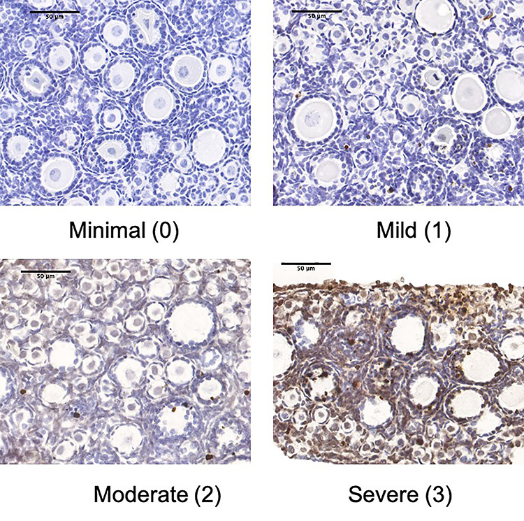
Phthalates are a group of chemicals used as additives in various consumer products, medical equipment, and personal care products. Phthalates and their metabolites are consistently detected in humans, indicating widespread and continuous exposure to multiple phthalates. Thus, environmentally relevant mixtures of phthalates and phthalate metabolites were investigated to determine the effects of phthalates on the function of the ovary during the neonatal period of development. Neonatal ovaries from CD-1 mice were cultured with dimethyl sulphoxide (DMSO; vehicle control), phthalate mixture (0.1-100 μg/mL), or phthalate metabolite mixture (0.1-100 μg/mL). The phthalate mixture was composed of 35% diethyl phthalate, 21% di(2-ethylhexyl) phthalate, 15% dibutyl phthalate, 15% diisononyl phthalate, 8% diisobutyl phthalate, and 5% benzylbutyl phthalate. The phthalate metabolite mixture was composed of 37% monoethyl phthalate, 19% mono(2-ethylhexyl) phthalate, 15% monobutyl phthalate, 10% monoisononyl phthalate, 10% monoisobutyl phthalate, and 8% monobenzyl phthalate. After 96 h of culture, ovaries were harvested for histological analysis of folliculogenesis, gene expression analysis of cell cycle and apoptosis regulators, and immune staining for cell proliferation and apoptosis. The metabolite mixture significantly decreased the number and percentage of abnormal follicles (100 μg/mL) compared to controls. The metabolite mixture also significantly increased the expression of cell cycle inhibitors (100 μg/mL) and the antiapoptotic factor Bcl2l10 (10 μg/mL) compared to controls. The phthalate mixture did not significantly alter gene expression or follicle counts, but ovaries exposed to the phthalate mixture (0.1 μg/mL) exhibited marginally significantly increased apoptosis as revealed by DNA fragmentation staining. Overall, these data show that parent phthalates and phthalate metabolites differentially impact ovarian function.

Keywords: apoptosis; cell cycle; endocrine disruption; folliculogenesis; mixtures; neonatal ovary; ovary; phthalates; toxicology.


Figures

Figure 1
Figure 1
A representative image of a hematoxylin and eosin-stained section. The labeled follicles in the inset are representatives of primordial follicles (1), primary follicles (2), preantral follicles (3), and abnormal (atretic) follicles (4).
Figure 2
Figure 2
Effects of the phthalate mixture and metabolite mixture on early folliculogenesis. Neonatal ovaries were isolated from PND 4 mice and treated with vehicle (DMSO), phthalate mixture (PM), or metabolite mixture (MM) (0.1–100 μg/mL) for 96 h. Ovaries were collected and processed for histological evaluation of follicle counts. The figure shows the total number of follicles and percentage of follicles at each stage of folliculogenesis from ovaries treated with the PM (A, B, respectively) and MM (C, D, respectively). Graphs represent mean ± SEM from 3–5 separate replicate experiments. Asterisks (*) indicate significant differences from the control (P ≤ 0.05); caret (^) indicates borderline significance compared to control (P ≤ 0.10).
Figure 3
Figure 3
Effects of the phthalate mixture and metabolite mixture on the expression of cell cycle regulators. Neonatal ovaries were isolated from PND 4 mice and treated with vehicle (DMSO), phthalate mixture, or metabolite mixture (0.1–100 μg/mL) for 96 h. Ovaries were collected and subjected to qPCR. Relative fold changes of each gene normalized to Rn18s are shown. Graphs represent mean ± SEM from 3–8 separate replicate experiments (n = 3 ovaries/treatment/experiment). Asterisks (*) indicate significant differences from the control (P ≤ 0.05); caret (^) indicates borderline significance compared to control (P ≤ 0.10).
Figure 4
Figure 4
Effects of the phthalate mixture and the metabolite mixture on the expression of apoptosis regulators. Neonatal ovaries were isolated from PND 4 mice and treated with vehicle (DMSO), phthalate mixture, or metabolite mixture (0.1–100 μg/mL) for 96 h. Ovaries were collected and subjected to qPCR. Relative fold changes of each gene normalized to Rn18s are shown. Graphs represent mean ± SEM from 3–8 separate experiments (n = 3 ovaries/treatment/experiment). Asterisks (*) indicate significant differences from the control (P ≤ 0.05); caret (^) indicates borderline significance compared to control (P ≤ 0.10).
Figure 5
Figure 5
Effects of the phthalate mixture and the metabolite mixture on Ki67 staining. Neonatal ovaries were isolated from PND 4 mice and treated with vehicle (DMSO), phthalate mixture, or metabolite mixture (0.1–100 μg/mL) for 96 h. Ovaries were collected and processed for immunohistochemistry of Ki67. Representative image of Ki67 staining shows staining primarily occurred in granulosa cells. The percent of positively stained pixels was determined using ImageJ. The graph represents mean ± SEM from 3–5 separate replicate experiments, except for 100 μg/mL MM, which had n = 2 because of problems with fixation.
Figure 6
Figure 6
Effects of the phthalate mixture and the metabolite mixture on apoptosis staining. Neonatal ovaries were isolated from PND 4 mice and treated with vehicle (DMSO), phthalate mixture, or metabolite mixture (0.1–100 μg/mL) for 96 h. Ovaries were collected and processed for TUNEL staining. Images were scored according to their level of staining, with minimal staining scored as 0, mild as 1, moderate as 2, and severe as 3.

References

    1. Woodruff TJ, Carlson A, Schwartz JM, Giudice LC. Proceedings of the summit on environmental challenges to reproductive health and fertility: Executive summary. Fertil Steril 2008; 89:281–300. - PMC - PubMed
    1. Marques-Pinto A, Carvalho D. Human infertility: Are endocrine disruptors to blame? Endocr Connect 2013; 2:R15–R29. - PMC - PubMed
    1. Koniecki D, Wang R, Moody RP, Zhu J. Phthalates in cosmetic and personal care products: Concentrations and possible dermal exposure. Environ Res 2011; 111:329–336. - PubMed
    1. Craig ZR, Ziv-Gal A. Pretty good or pretty bad? The ovary and chemicals in personal care products. Toxicol Sci 2018; 162:349–360. - PubMed
    1. Heudorf U, Mersch-Sundermann V, Angerer J. Phthalates: Toxicology and exposure. Int J Hyg Environ Health 2007; 210:623–634. - PubMed

Publication types