. 2021 Apr 29;137(17):2337-2346.
doi: 10.1182/blood.2020009499.

T-cell activation profiles distinguish hemophagocytic lymphohistiocytosis and early sepsis

Affiliations

T-cell activation profiles distinguish hemophagocytic lymphohistiocytosis and early sepsis

Vandana Chaturvedi et al. Blood. .

Abstract

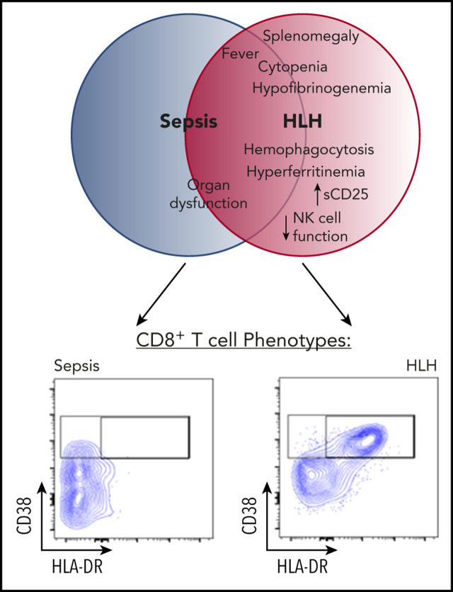
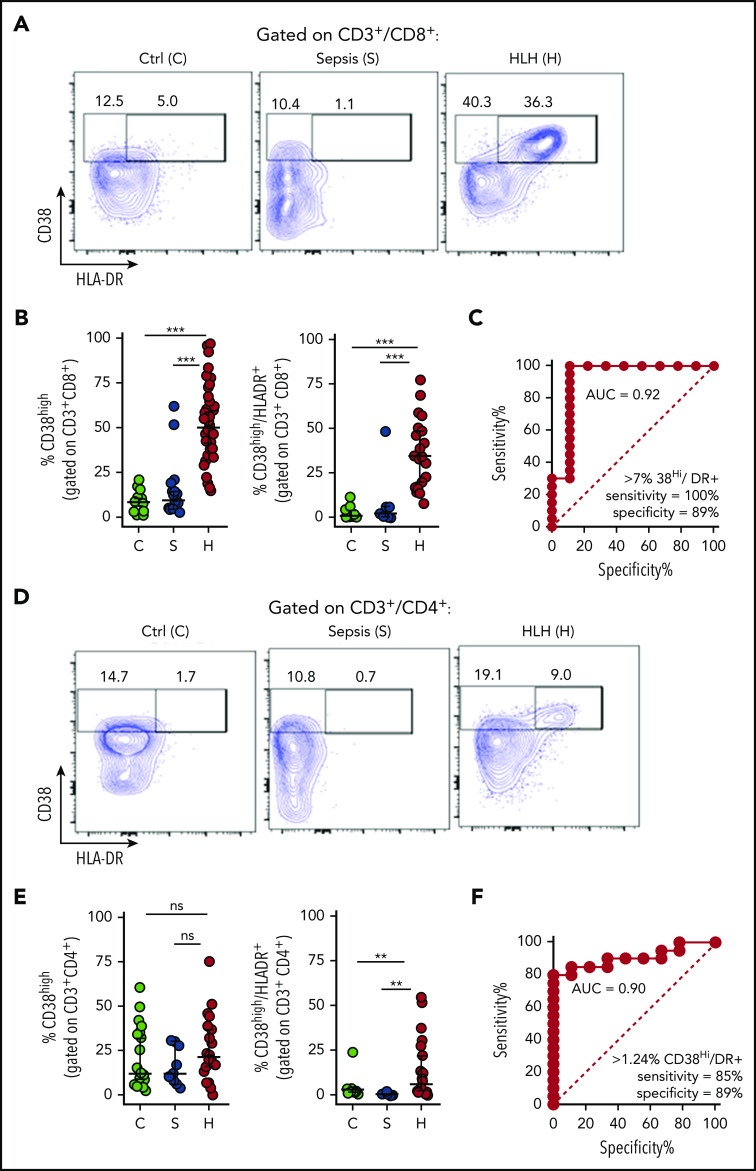
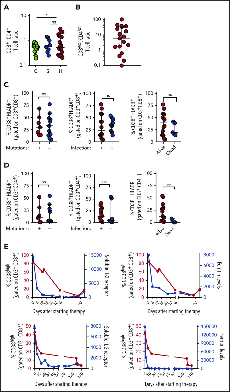
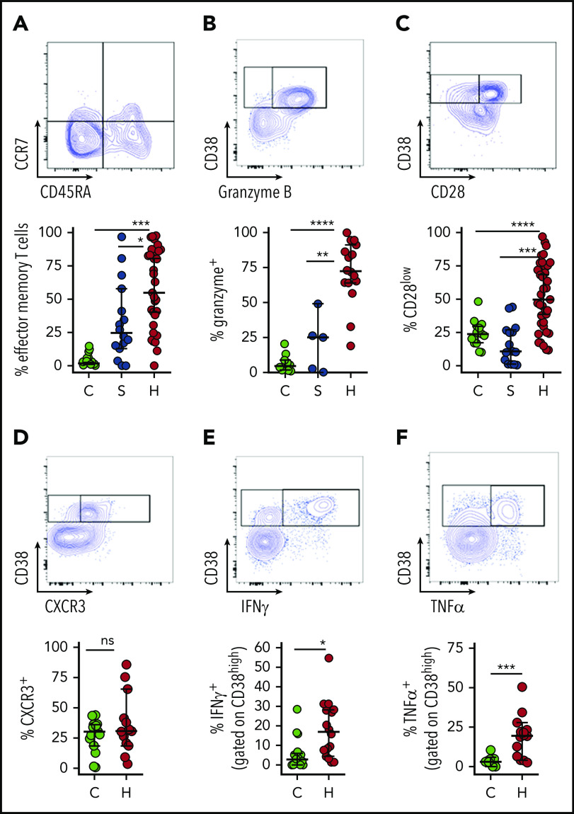
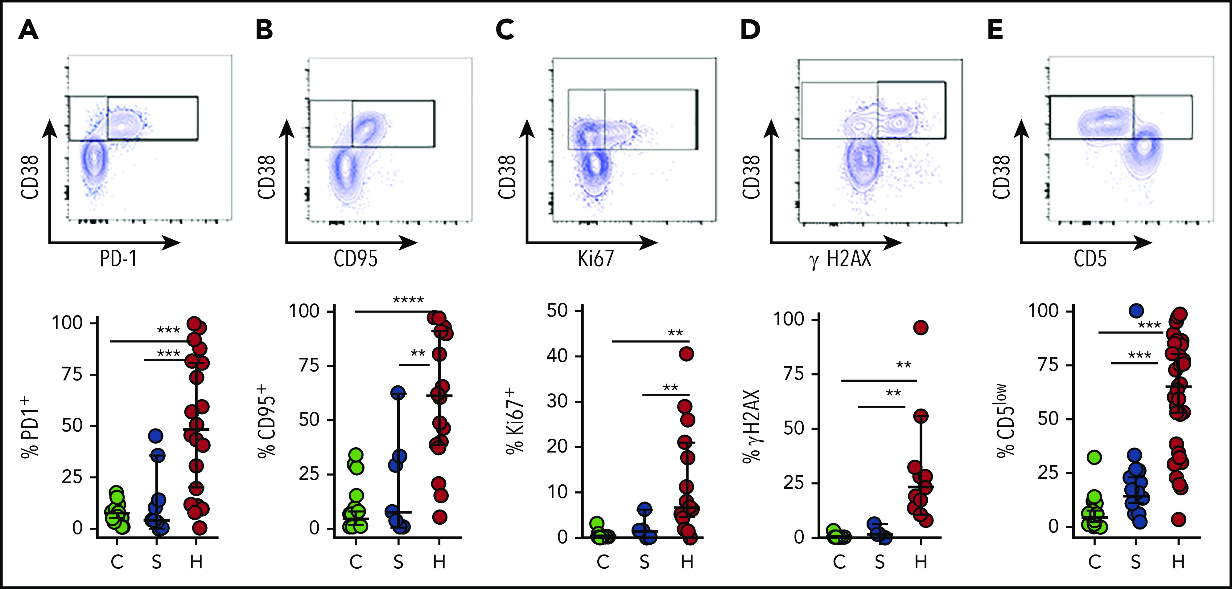
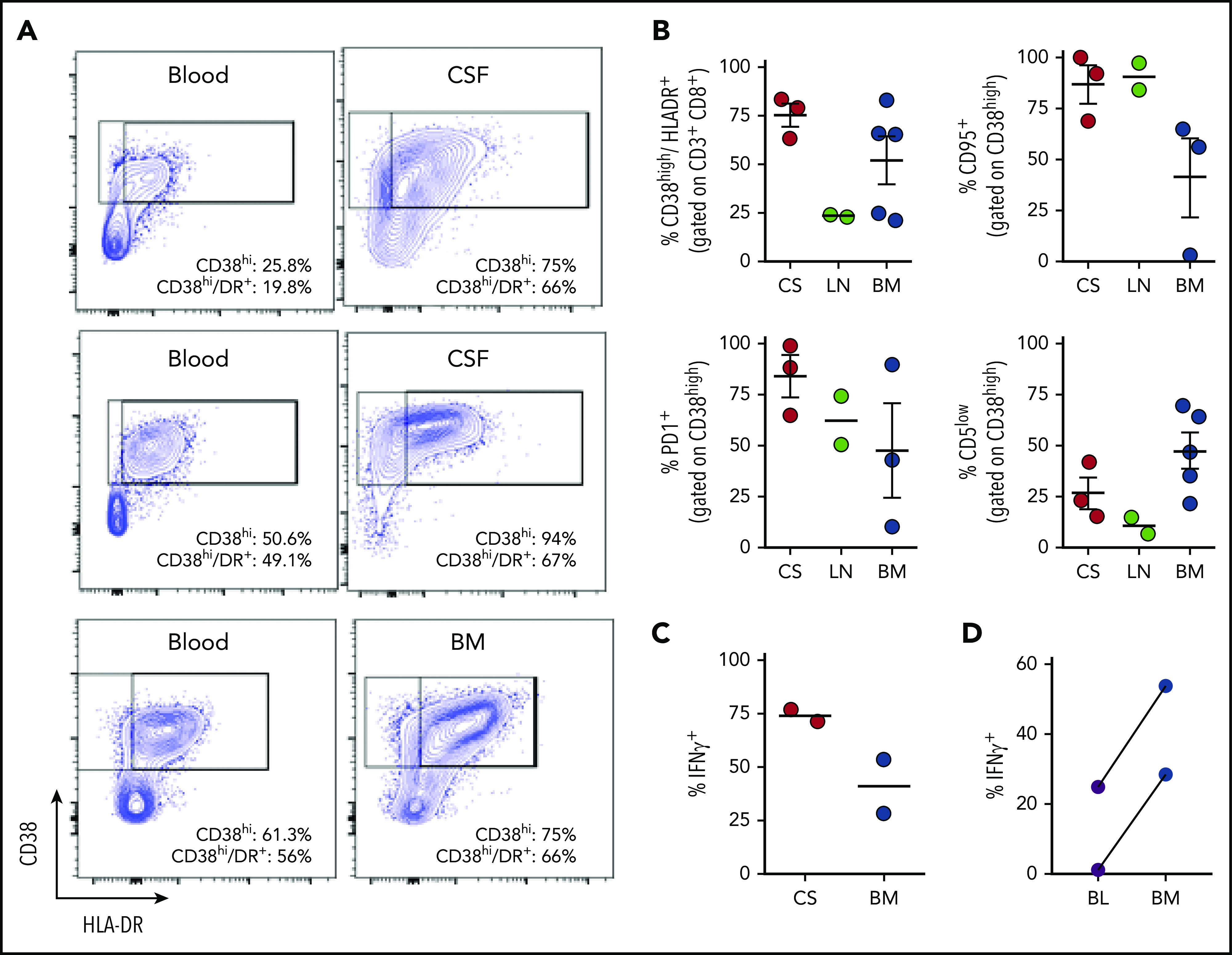
Hemophagocytic lymphohistiocytosis (HLH) is a fatal disorder of immune hyperactivation that has been described as a cytokine storm. Sepsis due to known or suspected infection has also been viewed as a cytokine storm. Although clinical similarities between these syndromes suggest similar immunopathology and may create diagnostic uncertainty, distinguishing them is critical as treatments are widely divergent. We examined T-cell profiles from children with either HLH or sepsis and found that HLH is characterized by acute T-cell activation, in clear contrast to sepsis. Activated T cells in patients with HLH were characterized as CD38high/HLA-DR+ effector cells, with activation of CD8+ T cells being most pronounced. Activated T cells were type 1 polarized, proliferative, and displayed evidence of recent and persistent activation. Circulating activated T cells appeared to be broadly characteristic of HLH, as they were seen in children with and without genetic lesions or identifiable infections and resolved with conventional treatment of HLH. Furthermore, we observed even greater activation and type 1 polarization in tissue-infiltrating T cells, described here for the first time in a series of patients with HLH. Finally, we observed that a threshold of >7% CD38high/HLA-DR+ cells among CD8+ T cells had strong positive and negative predictive value for distinguishing HLH from early sepsis or healthy controls. We conclude that the cytokine storm of HLH is marked by distinctive T-cell activation whereas early sepsis is not, and that these 2 syndromes can be readily distinguished by T-cell phenotypes.


Conflict of interest statement

Conflict-of-interest disclosure: M.B.J., M.L.H., and C.E.A. have served as consultants for Sobi. M.L.H. serves on a data safety monitoring committee for Novimmune. The remaining authors declare no competing financial interests.

Figures

None
Graphical abstract
Figure 1.
Figure 1.
Peripheral blood T-cell activation status readily distinguishes patients with HLH and sepsis. (A-B) Representative flow cytometric plots and cumulative data comparing the frequency of CD38high or CD38high/HLA-DR+ (double-positive) CD8+ T cells in pediatric controls (C), patients with sepsis (S), or patients with HLH (H). (C) ROC analysis of CD38high/HLA-DR+ (double-positive) CD8+ T cells, comparing HLH and sepsis. (D-E) Representative FACS plots and cumulative data comparing the frequency of CD38high or CD38high/HLA-DR+ (double-positive) CD4+ T cells in pediatric controls, patients with sepsis, or patients with HLH. (F) ROC analysis of CD38high/HLA-DR+ (double-positive) CD4+ T cells, comparing HLH and sepsis. Data are representative of 27 pediatric controls, 9 to 19 patients with sepsis and 19 to 43 patients with HLH. Error bars represent median with 95% CI. Differences between indicated groups were calculated using the unpaired Student t test. **P < .01; ***P < .001. Ctrl, control; ns, not significant.
Figure 2.
Figure 2.
Increased frequency of CD38high/HLA-DR+ CD8+ T cells is a robust marker of active HLH, irrespective of clinical context. (A) CD8:CD4 ratio is modestly elevated in patients with HLH, compared with controls, but not patients with sepsis. (B) Ratio of CD38high/HLA-DR+ CD8+ to CD38high/HLA-DR+ CD4+ T cells in individual patients with HLH. (C-D) Frequency of CD38high/HLA-DR+ CD8+ or CD4+ T cells in patients with HLH is plotted, comparing presence of HLH-associated mutations, identifiable infection at diagnosis, or subsequent survival. (E) Frequency of CD38high CD8+ T cells is plotted with either sCD25 or ferritin across time in 2 patients receiving etoposide and dexamethasone for HLH. Data are representative of 27 pediatric controls, 9 patients with sepsis, and 18 to 22 patients with HLH. Error bars represent median with 95% CI. Differences between indicated groups were calculated using the unpaired Student t test. *P < .05.
Figure 3.
Figure 3.
Activated CD8+ T cells in patients with HLH are predominantly effector memory T cells with Tc1 and cytotoxic differentiation. Representative flow cytometry plots from patients with HLH and composite data comparing controls and patients with sepsis or HLH, showing (A) frequency effector T-cell differentiation (CD45RA CCR7) among CD38high/HLA-DR+ CD8+ T cells. As CD38+/DR+ T cells are largely absent from healthy controls and patients with sepsis, frequency shown is derived from gating on the top 10% of CD38-expressing CD8+ T cells. (B-C) Expression of CD28 or granzyme B in CD38high/HLA-DR+ CD8+ T cells is shown as in panel A. (D) Representative FACS plots and composite data showing CXCR3 expression in CD8+ CD38high T cells of controls (top 10% of CD38 expressors) and patients with HLH. (E-F) Representative flow cytometry plots and composite data showing IFN-γ and TNF-α production among CD8+CD38high cells in indicated populations after phorbol myristate acetate/ionomycin stimulation. Data are representative of 12 to 27 pediatric controls, 5 to 16 patients with sepsis, and 13 to 37 patients with HLH. Error bars represent median with 95% CI. Differences between indicated groups were calculated using the unpaired Student t test. *P < .05; **P < .01; ***P < .001; ****P < .0001.
Figure 4.
Figure 4.
CD38high/HLA-DR+ CD8+ T cells in patients with HLH are proliferative and display evidence of recent and persistent activation. (A-E) Representative flow cytometry plots from patients with HLH and composite data comparing controls and patients with sepsis or HLH, showing the indicated markers of activation, proliferation, and persistent stimulation (top of each panel). For controls and patients with sepsis, frequency shown is derived from gating on the top 10% of CD38-expressing CD8+ T cells, as CD38+/DR+ T cells are largely absent in these samples (bottom of each panel). Data are representative of 18 to 27 pediatric controls, 5 to 18 patients with sepsis, and 11 to 36 patients with HLH. Error bars represent median with 95% CI. Differences between indicated groups were calculated using the unpaired Student t test. **P < .01; ***P < .001; ****P < .0001.
Figure 5.
Figure 5.
Tissue-infiltrating CD8+ T cells in patients with HLH are highly activated, IFN-γ–producing T cells. (A) Representative FACS plots of CD8+ T cells comparing blood and CSF or bone marrow in samples obtained concurrently from 3 different patients. (B) Cumulative data for the indicated markers in samples of CSF (CS), lymph node (LN), or bone marrow (BM) from 10 patients with HLH. CD38high/HLA-DR+ cells are displayed as a percentage of CD3+/CD8+ cells, the other markers are displayed as a percentage of CD8/CD38high/HLA-DR+ cells. (C) Representative FACS plots and cumulative data of intracellular cytokine staining for IFN-γ, as a percentage of CD3+/CD8+ cells in either CSF or bone marrow. (D) Comparison of IFN-γ staining of CD8+/CD38high T cells after stimulation of concurrent peripheral blood (BL) and bone marrow samples from 2 patients.

Comment in

References

    1. Sriskandan S, Altmann DM. The immunology of sepsis. J Pathol. 2008;214(2):211-223. - PubMed
    1. Rothman AL. Immunity to dengue virus: a tale of original antigenic sin and tropical cytokine storms. Nat Rev Immunol. 2011;11(8):532-543. - PubMed
    1. Mehta P, McAuley DF, Brown M, Sanchez E, Tattersall RS, Manson JJ; HLH Across Speciality Collaboration, UK . COVID-19: consider cytokine storm syndromes and immunosuppression. Lancet. 2020;395(10229):1033-1034. - PMC - PubMed
    1. Ferrara JL, Abhyankar S, Gilliland DG. Cytokine storm of graft-versus-host disease: a critical effector role for interleukin-1. Transplant Proc. 1993;25(1 pt 2):1216-1217. - PubMed
    1. Suntharalingam G, Perry MR, Ward S, et al. . Cytokine storm in a phase 1 trial of the anti-CD28 monoclonal antibody TGN1412. N Engl J Med. 2006;355(10):1018-1028. - PubMed

Publication types

MeSH terms