. 2021 Feb 25;137(8):1061-1071.
doi: 10.1182/blood.2020008762.

Antibody-induced procoagulant platelets in severe COVID-19 infection

Affiliations

Antibody-induced procoagulant platelets in severe COVID-19 infection

Karina Althaus et al. Blood. .

Abstract

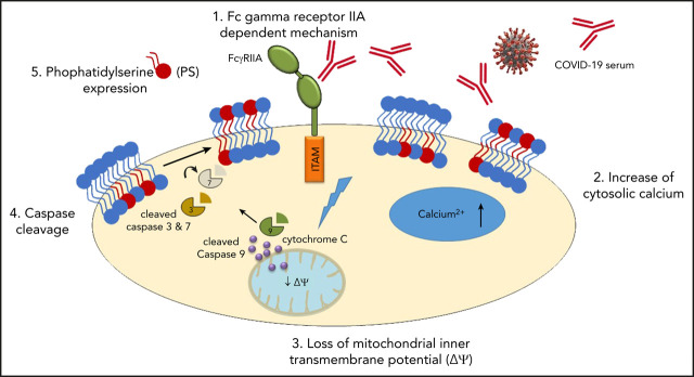
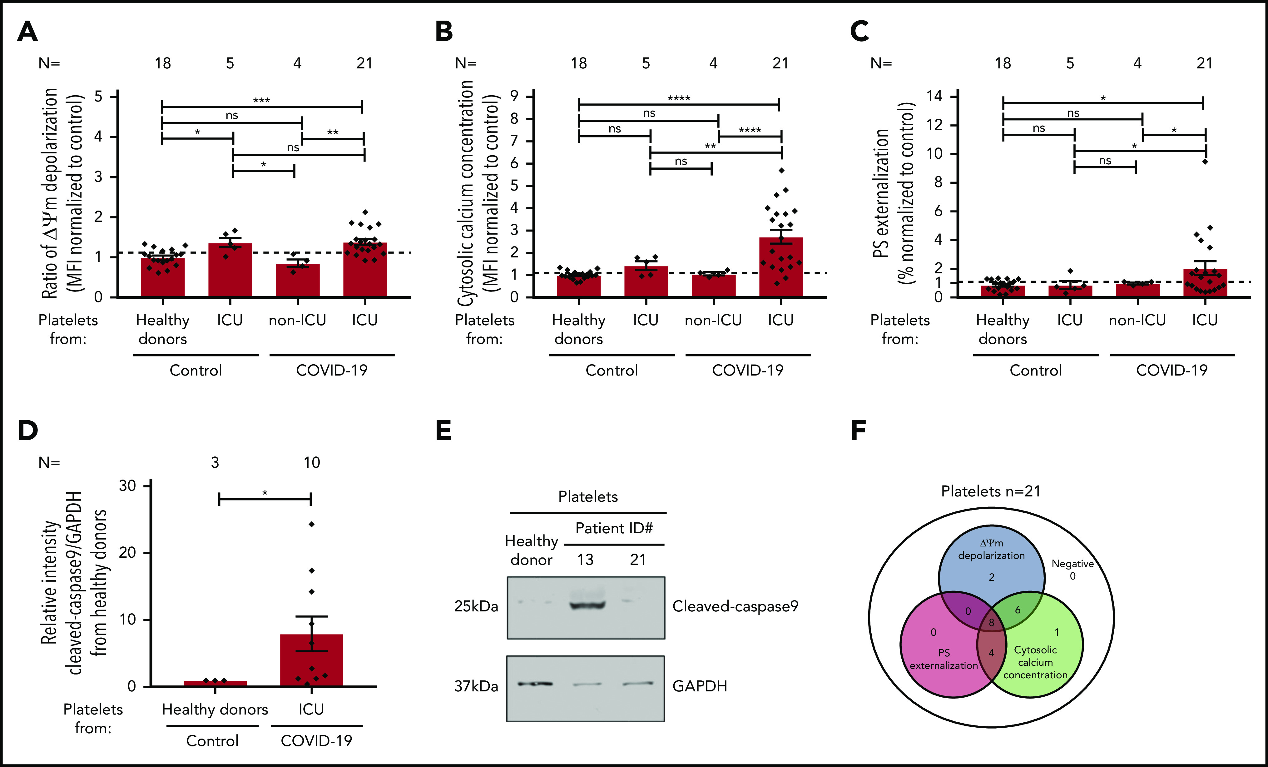
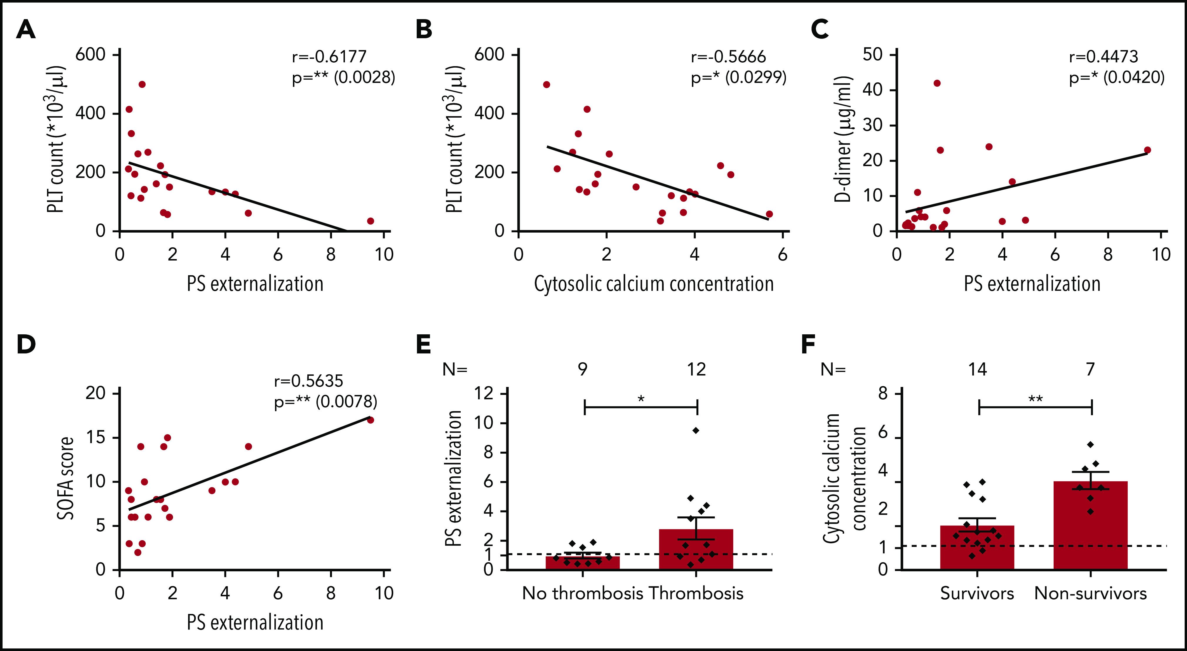
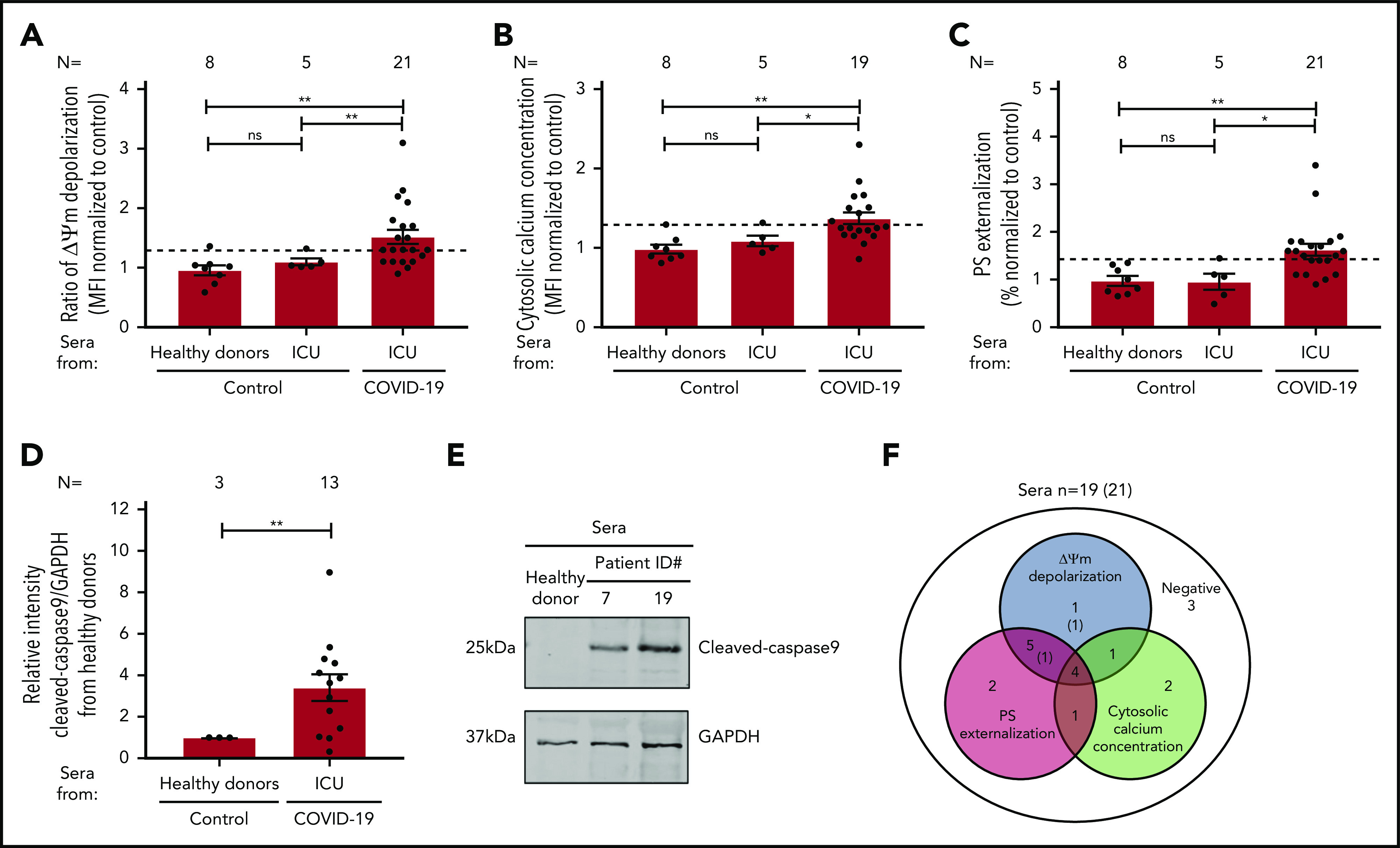
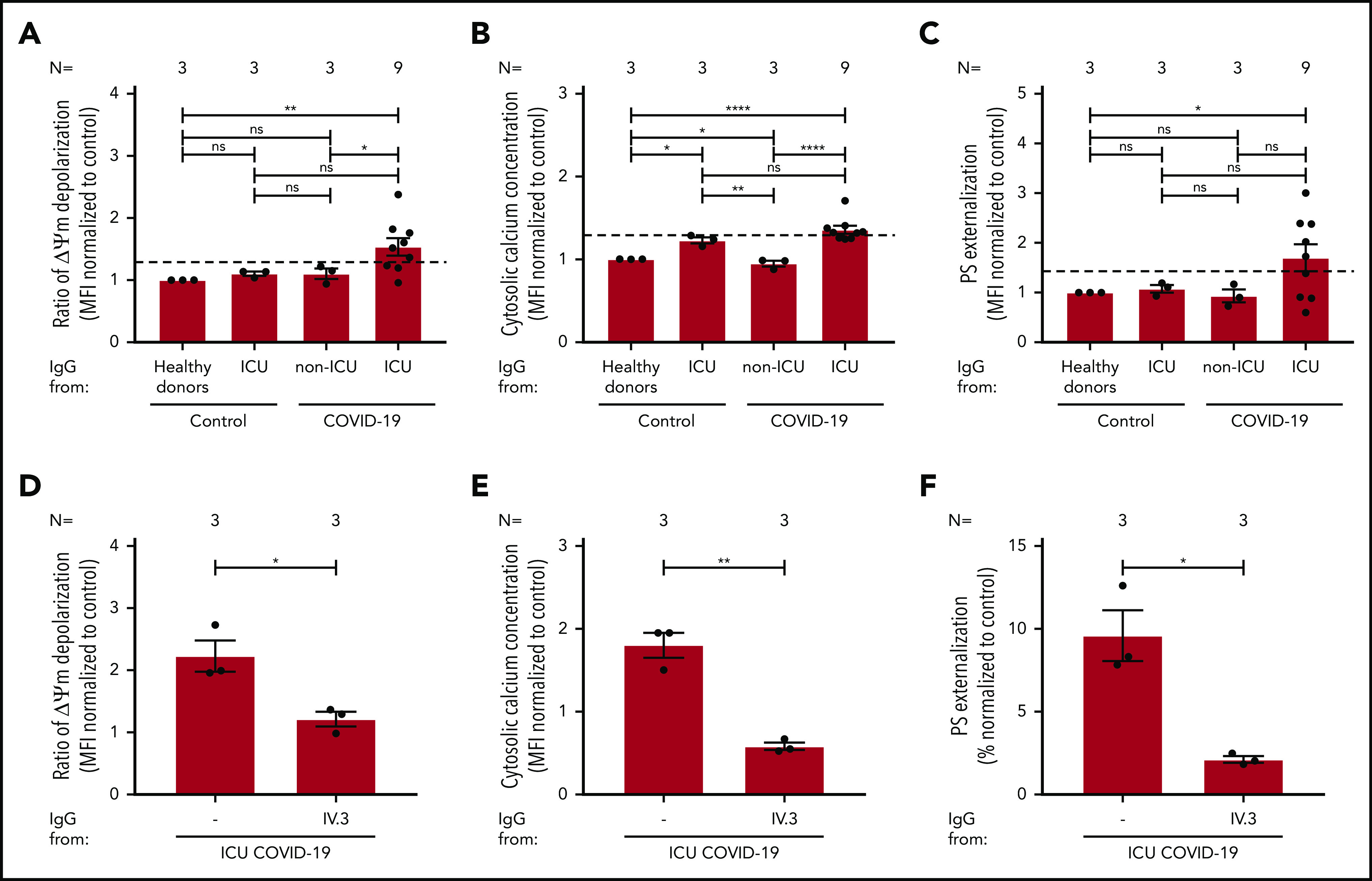
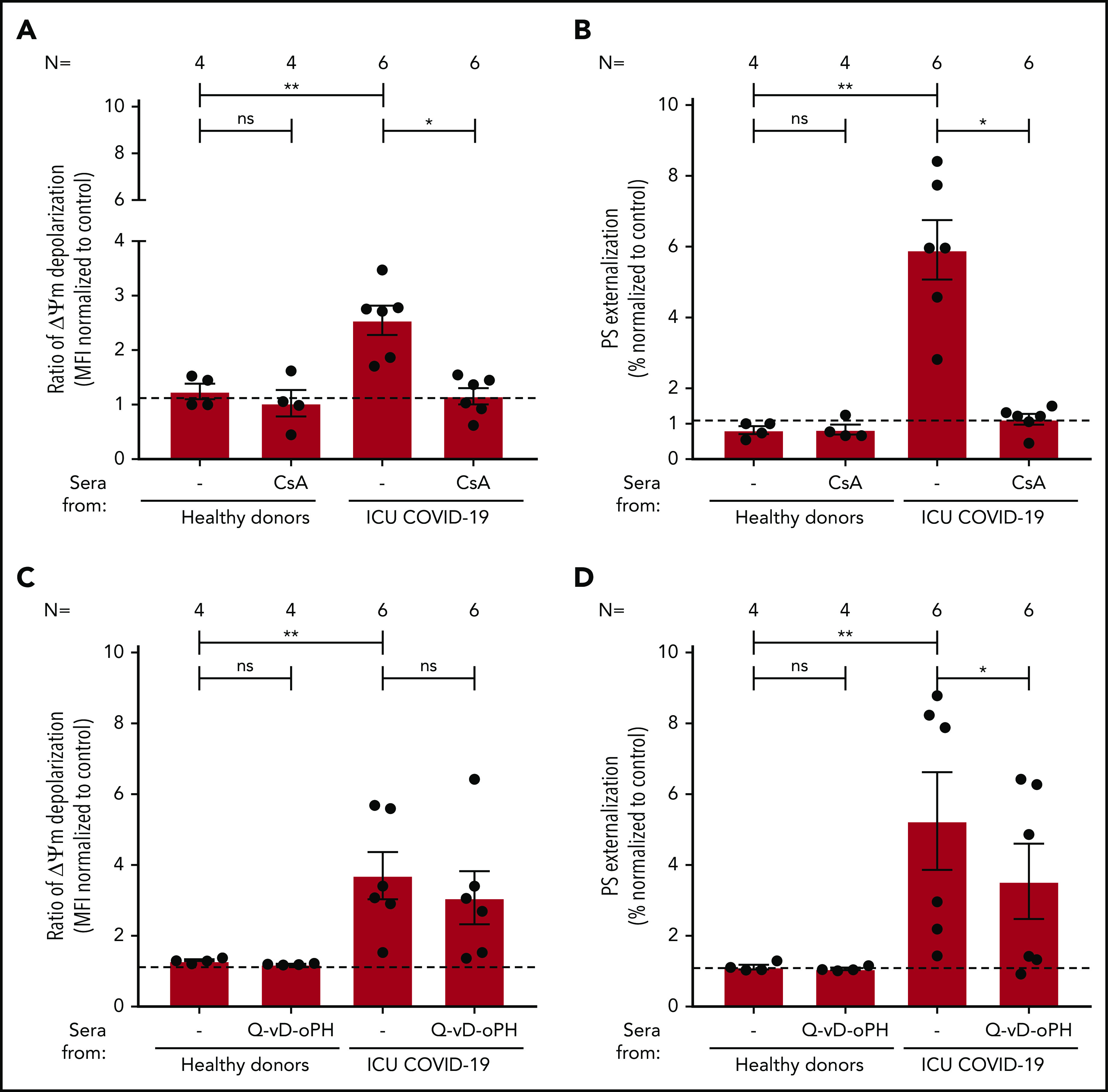
The pathophysiology of COVID-19-associated thrombosis seems to be multifactorial. We hypothesized that COVID-19 is accompanied by procoagulant platelets with subsequent alteration of the coagulation system. We investigated depolarization of mitochondrial inner transmembrane potential (ΔΨm), cytosolic calcium (Ca2+) concentration, and phosphatidylserine (PS) externalization. Platelets from COVID-19 patients in the intensive care unit (ICU; n = 21) showed higher ΔΨm depolarization, cytosolic Ca2+, and PS externalization compared with healthy controls (n = 18) and non-ICU COVID-19 patients (n = 4). Moreover, significant higher cytosolic Ca2+ and PS were observed compared with a septic ICU control group (ICU control; n = 5). In the ICU control group, cytosolic Ca2+ and PS externalization were comparable with healthy controls, with an increase in ΔΨm depolarization. Sera from COVID-19 patients in the ICU induced a significant increase in apoptosis markers (ΔΨm depolarization, cytosolic Ca2+, and PS externalization) compared with healthy volunteers and septic ICU controls. Interestingly, immunoglobulin G fractions from COVID-19 patients induced an Fcγ receptor IIA-dependent platelet apoptosis (ΔΨm depolarization, cytosolic Ca2+, and PS externalization). Enhanced PS externalization in platelets from COVID-19 patients in the ICU was associated with increased sequential organ failure assessment score (r = 0.5635) and D-dimer (r = 0.4473). Most importantly, patients with thrombosis had significantly higher PS externalization compared with those without. The strong correlations between markers for apoptosic and procoagulant platelets and D-dimer levels, as well as the incidence of thrombosis, may indicate that antibody-mediated procoagulant platelets potentially contributes to sustained increased thromboembolic risk in ICU COVID-19 patients.


Conflict of interest statement

Conflict-of-interest disclosure: The authors declare no competing financial interests.

Figures

None
Graphical abstract
Figure 1.
Figure 1.
Platelet apoptosis in COVID-19 patients. (A-C) Changes in apoptosis pathways were analyzed by assessing the depolarization of the ΔΨm (A), cytosolic calcium concentration (B), and PS externalization (C) in platelets from COVID-19 patients in the ICU or COVID-19 patients not in the ICU, as well as ICU non–COVID-19 patients (control group) and healthy donors, respectively. (D) Quantification of cleaved-caspase 9 level in platelets from COVID-19 patients in the ICU normalized to healthy donors. (E) Representative western blot showing GAPDH and cleaved-caspase 9 proteins in platelets from COVID-19 patients in the ICU. Protein bands were detected with the infrared imaging system (Odyssey, LI.COR, Lincoln, NE). (F) Diagram indicating the number of COVID-19 patients in the ICU positive for each apoptotic parameter: ΔΨm depolarization, cytosolic calcium concentration, and PS externalization. Data are presented as mean ± standard error mean (SEM) of the measured fold increase compared with control. Not significant, *P < .05, **P < .01, ***P < .001, ****P < .0001. The number of patients and healthy donors tested is reported in each graphic. Dashed lines represent the cutoffs determined from healthy donors as mean of fold increase (FI) + 2× SEM.
Figure 2.
Figure 2.
Association among platelet apoptosis and clinical biomarkers, thromboembolic complications, and mortality. The correlations between platelet apoptosis parameters and PLT count, as well as D-dimer and SOFA score measured at the same day of platelet testing, were assessed. (A-B) An association was observed between PLT count and PS externalization (A) and cytosolic calcium concentration (B). (C) Moreover, a significant correlation was detected for D-dimer and PS externalization. (D) The clinical relevance of PS externalization was assessed using the SOFA score and revealed a significant positive correlation. Pearson’s correlation coefficients were calculated and are shown in the panels. (E-F) The PS externalization (E) and the cytosolic calcium concentration (F) were determined and compared between COVID-19 patients in the ICU depending on the incidence of thromboembolic complications and mortality, respectively. Data are presented as mean of the measured FI compared with control. Not significant, *P < .05, **P < .01, ***P < .001, ****P < .0001. The number of patients and healthy donors tested is reported in each graphic. Dashed lines represent the cutoffs determined from healthy donors as mean of FI + 2× SEM.
Figure 3.
Figure 3.
Impact of sera from COVID-19 patients on platelet apoptosis. (A-C) Changes in apoptosis pathways induced by sera from COVID-19 patients in the ICU, non–COVID-19 patients in the ICU (control group), and healthy donors were investigated by assessing the depolarization of the ΔΨm (A), cytosolic calcium concentration (B) (n = 19 because of the lack of biomaterial for 2 patients), and PS externalization (C). (D) Quantification of cleaved caspase 9 level in platelets from healthy donors after incubation with patient sera, normalized to sera from healthy donors. (E) Representative western blot of GAPDH and cleaved caspase 9 proteins. Protein bands were detected with the infrared imaging system (Odyssey, LI.COR). (F) Diagram indicating the number of COVID-19 patients in the ICU positive for each apoptotic parameter: ΔΨm depolarization, cytosolic calcium concentration, and PS externalization. The 2 sera tested only for ΔΨm depolarization and PS externalization are in parentheses. Data are presented as mean ± SEM of the measured FI compared with control. Not significant, *P < .05, **P < .01, ***P < .001, ****P < .0001. The number of sera tested is reported in each graphic. Dashed lines represent the cutoffs determined testing sera from healthy donors as mean of FI + 2× SEM.
Figure 4.
Figure 4.
IgG fractions from COVID-19 patients in the ICU induce platelet apoptosis via crosslinking FcγRIIA. (A-C) Changes in apoptosis pathways induced by IgG fraction from COVID-19 patients in the ICU or COVID-19 patients not in the ICU, as well as non–COVID-19 patients in the ICU (control group) and healthy donors, were analyzed by assessing the depolarization of the ΔΨm (A), cytosolic calcium concentration (B), and PS externalization (C) in platelets from 3 different healthy donors. (D-F) The same assays were performed in the presence of the IV.3 monoclonal antibody (mAb) to block FcγRIIA signaling. Each dot represents 2 experiments with platelets from 2 different donors. Data are presented as mean ± SEM of the measured FI compared with control. Not significant, *P < .05, **P < .01, ***P < .001, ****P < .0001. The number of sera tested is reported in each graphic.
Figure 5.
Figure 5.
The impact of CsA and Q-VD-OPh on antibody-mediated procoagulant platelets. (A-B) CsA and Q-VD-OPh inhibition of COVID-19–induced ΔΨ depolarization (A) and PS externalization (B). (C-D) CsA significantly inhibits the depolarization of ΔΨm and PS expression in COVID-19–positive patients. Q-VD-OPh was not able to inhibit ΔΨm depolarization (C) but significantly reduced PS externalization (D). Data are presented as mean ± SEM of the measured FI compared with control. Not significant, *P < .05, **P < .01, ***P < .001, ****P < .0001. The number of sera tested is reported in each graphic.

Comment in

References

    1. Violi F, Pastori D, Cangemi R, Pignatelli P, Loffredo L. Hypercoagulation and antithrombotic treatment in coronavirus 2019: a new challenge. Thromb Haemost. 2020;120(6):949-956. - PMC - PubMed
    1. Levi M, Thachil J, Iba T, Levy JH. Coagulation abnormalities and thrombosis in patients with COVID-19. Lancet Haematol. 2020;7(6):e438-e440. - PMC - PubMed
    1. Connors JM, Levy JH. COVID-19 and its implications for thrombosis and anticoagulation. Blood. 2020;135(23):2033-2040. - PMC - PubMed
    1. Klok FA, Kruip MJHA, van der Meer NJM, et al. . Confirmation of the high cumulative incidence of thrombotic complications in critically ill ICU patients with COVID-19: an updated analysis. Thromb Res. 2020;191:148-150. - PMC - PubMed
    1. Heemskerk JW, Mattheij NJ, Cosemans JM. Platelet-based coagulation: different populations, different functions. J Thromb Haemost. 2013;11(1):2-16. - PubMed

Publication types

MeSH terms