Review
. 2021 Feb 4;79(2):ftaa078.
doi: 10.1093/femspd/ftaa078.

Got mutants? How advances in chlamydial genetics have furthered the study of effector proteins

Affiliations
Review

Got mutants? How advances in chlamydial genetics have furthered the study of effector proteins

Shelby E Andersen et al. Pathog Dis. .

Abstract

Chlamydia trachomatis is the leading cause of infectious blindness and a sexually transmitted infection. All chlamydiae are obligate intracellular bacteria that replicate within a membrane-bound vacuole termed the inclusion. From the confines of the inclusion, the bacteria must interact with many host organelles to acquire key nutrients necessary for replication, all while promoting host cell viability and subverting host defense mechanisms. To achieve these feats, C. trachomatis delivers an arsenal of virulence factors into the eukaryotic cell via a type 3 secretion system (T3SS) that facilitates invasion, manipulation of host vesicular trafficking, subversion of host defense mechanisms and promotes bacteria egress at the conclusion of the developmental cycle. A subset of these proteins intercalate into the inclusion and are thus referred to as inclusion membrane proteins. Whereas others, referred to as conventional T3SS effectors, are released into the host cell where they localize to various eukaryotic organelles or remain in the cytosol. Here, we discuss the functions of T3SS effector proteins with a focus on how advances in chlamydial genetics have facilitated the identification and molecular characterization of these important factors.

Keywords: Chlamydia; effector; genetics; inclusion; inclusion membrane protein; type III secretion.


Figures

Figure 1.
Figure 1.
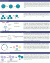
The intracellular life cycle of Chlamydia. Contact between the EB and the host cell triggers delivery of pre-packaged effector proteins that trigger cytoskeletal and membrane remodeling events to promote invasion. The nascent inclusion avoids fusion with lysosomes and traffics along microtubules to the MTOC. EBs convert to RBs and replication ensues via polarized cell division. Throughout the infection cycle, additional effector proteins are delivered into the host cell or the inclusion membrane to mediate interactions with various host organelles. RBs undergo asynchronous conversion to EBs and at the conclusion of the developmental cycle, EBs are released by extrusion or lysis.
Figure 2.
Figure 2.
Advances in chlamydial genetics. (A) Initial experiments to transform chlamydiae used electroporation; however, (B)C. trachomatis serovar L2 is routinely transformed via chemical transformation with calcium chloride. Using the endogenous L2 plasmid fused to an E. coli plasmid, a C. trachomatis L2 shuttle vector was developed. This plasmid possesses a GFP fluorescent marker, antibiotic selection marker (bla), an E. coli origin of replication and a multiple cloning site (MCS). The shuttle vector is routinely used to express epitope-tagged effector proteins in C. trachomatis L2. (C)The group II intron (TargeTron) approach enables site-specific gene disruption via LtrA. LtrA reverse transcribes and splices the intron into the target site in the recipient's DNA, resulting in insertional inactivation of the target. (D) Site-specific mutagenesis via fluorescence-reported allelic exchange mutagenesis (FRAEM) uses the shuttle vector pSU6 to disrupt the target gene of interest. In the absence of tetracycline, pSU6 behaves as a suicide vector. (E) The Himar1 transposase randomly inserts between T/A nucleotides, resulting in non-specific gene inactivation.(F) Mutants generated via any of the aforementioned techniques can be complemented using pBomb3, pBomb4 or pSU6.
Figure 3.
Figure 3.
TarP and TmeA, mediators of host cell invasion. TmeA and TarP are pre-packaged in EBs and are delivered into the host cell to promote invasion. TarP is phosphorylated upon entry into the host cell where it binds to Rac GEFs (SOS and VAV2) to activate the small GTPase Rac, which results in ARP2/3-dependent actin branching via complex activation by the NPF WAVE2. TarP can also directly bundle and polymerize actin. Focal adhesion proteins, FAK and vinculin, are also bound by TarP and may serve to promote cell adherence to the extracellular matrix. TmeA binds to the NPF N-WASP to promote ARP2/3-dependent actin branching and filopodia capture of EBs. TmeA also binds AHNAK, which may serve a post-invasion role.
Figure 4.
Figure 4.
Interactions with centrosomes and subversion of the cytoskeleton by Inc proteins. The Inc protein CT850 interacts with dynein light-chain DYNLT1 to facilitate inclusion positioning at the MTOC. CT288 binds to the centrosomal protein CCDC146. InaC is a multifunctional Inc that interacts with ARF GTPases to control Golgi positioning at the inclusion. InaC is also important for F-actin recruitment to the inclusion. CT228 and MraC inhibit or promote extrusion, respectively, through regulation of MLC2 phosphorylation state.
Figure 5.
Figure 5.
Manipulation of host vesicular trafficking by Inc and cT3SS effector proteins. CpoS interacts with Rab GTPases to recruit the transferrin receptor to the inclusion. IncV, through interactions with VAPs, functions to tether the inclusion to the ER. CteG localizes to the Golgi apparatus and plasma membrane and may be involved in regulating trafficking. IncA is involved with homotypic inclusion fusion and interacts with several VAMPs.

References

    1. Abdelrahman YM, Belland RJ. The chlamydial developmental cycle. FEMS Microbiol Rev. 2005;29:949–59. - PubMed
    1. Abdelrahman YM, Ouellette SP, Belland RJet al. . Polarized cell division of Chlamydia trachomatis. PLoS Pathog. 2016;12:1–20. - PMC - PubMed
    1. Agaisse H, Derré I.. A C. trachomatis cloning vector and the generation of C. trachomatis strains expressing fluorescent proteins under the control of a C. trachomatis promoter. PLoS One. 2013;8:e57090, DOI: 10.1371/journal.pone.0057090. - PMC - PubMed
    1. Agaisse H, Derré I. Expression of the effector protein IncD in Chlamydia trachomatis mediates recruitment of the lipid transfer protein CERT and the endoplasmic reticulum-resident protein VAPB to the inclusion membrane. Infect Immun. 2014;82:2037–47. - PMC - PubMed
    1. Al-Zeer MA, Al-Younes HM, Kerr Met al. . Chlamydia trachomatis remodels stable microtubules to coordinate Golgi stack recruitment to the chlamydial inclusion surface. Mol Microbiol. 2014;94:1285–97. - PubMed

Publication types

MeSH terms