. 2021 Jan 27;10(2):241.
doi: 10.3390/cells10020241.

Spermatogonia Loss Correlates with LAMA 1 Expression in Human Prepubertal Testes Stored for Fertility Preservation

Affiliations

Spermatogonia Loss Correlates with LAMA 1 Expression in Human Prepubertal Testes Stored for Fertility Preservation

Magdalena Kurek et al. Cells. .

Abstract

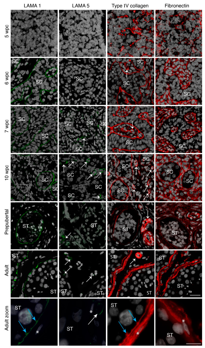
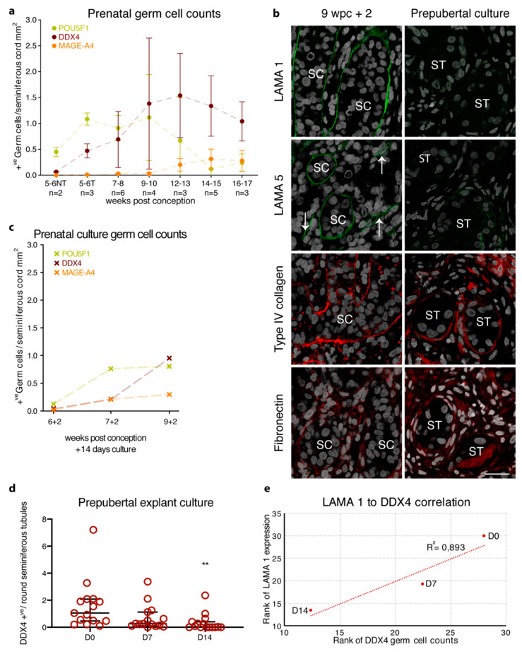
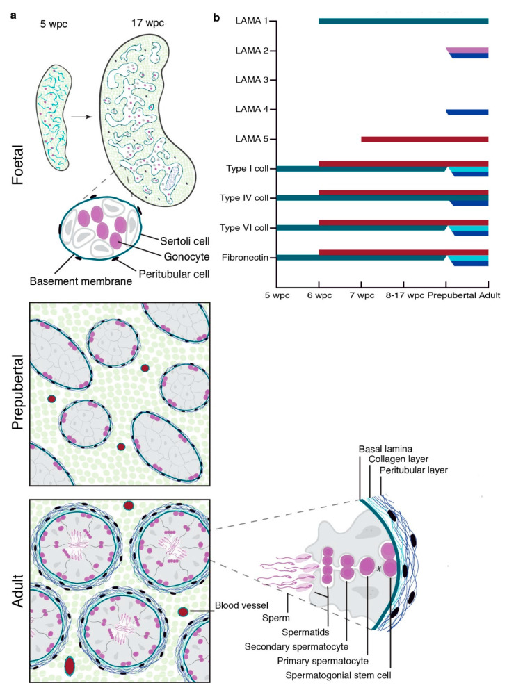
Fertility preservation for male childhood cancer survivors not yet capable of producing mature spermatozoa, relies on experimental approaches such as testicular explant culture. Although the first steps in somatic maturation can be observed in human testicular explant cultures, germ cell depletion is a common obstacle. Hence, understanding the spermatogonial stem cell (SSC) niche environment and in particular, specific components such as the seminiferous basement membrane (BM) will allow progression of testicular explant cultures. Here, we revealed that the seminiferous BM is established from 6 weeks post conception with the expression of laminin alpha 1 (LAMA 1) and type IV collagen, which persist as key components throughout development. With prepubertal testicular explant culture we found that seminiferous LAMA 1 expression is disrupted and depleted with culture time correlating with germ cell loss. These findings highlight the importance of LAMA 1 for the human SSC niche and its sensitivity to culture conditions.

Keywords: Sertoli cells; basal membrane; germ cells; infertility; late effects; seminiferous tubules; spermatogonia; stem cell niche.


Conflict of interest statement

The authors declare no conflict of interest.

Figures

Figure 1
Figure 1
Basement membrane expression of prenatal gonadal and postnatal testicular tissue. Laminin alpha 1 (LAMA 1) (green staining) expression is shown from 6 wpc onwards in the seminiferous basement membrane (BM), while LAMA 5 (green staining) can be observed only in the vasculature from 7 wpc onwards. Type IV collagen (red staining) and fibronectin (red staining) expression is noted from 5 wpc as a net-like structure in the seminiferous BM and also the interstitium in prenatal samples from 6 wpc onwards. Prenatal sample n = 27. Prepubertal testicular samples show LAMA 1 expression in the seminiferous BM while LAMA 5 can be observed in the vasculature. Type IV collagen and fibronectin is noted in the seminiferous BM, the vasculature and the interstitium. Prepubertal sample n = 16. Adult testicular samples show specialization with LAMA 1 expression in the innermost BM, type IV collagen expression throughout the entire BM, and fibronectin expression in the peritubular BM layer. Adult sample n = 3. Counterstain with DAPI (grey staining). SC depicts the seminiferous cord, ST depicts seminiferous tubules, white arrows indicate blood vessels, blue arrows show innermost BM protrusions between tubular cells, * indicate peritubular cells, scale bar: 25 μm, zoom scale bar: 10 µm.
Figure 2
Figure 2
Identification of cell clusters and single-cell transcription profiles of prenatal gonadal cells. (a) The t-distributed stochastic neighbour embedding (t-SNE) plot of germ and somatic cells, coloured by the identified cell types. FGC indicates foetal germ cells. (b) Single-cell expression profile for extra cellular matrix (ECM) protein coding genes exhibited on the t-SNE plot; gradient of grey, yellow, orange, and red indicates low to high expression.
Figure 3
Figure 3
Identification of cell clusters and single-cell transcription profiles of postnatal testicular cells. (a) The t-SNE plot of germ and somatic cells, coloured by the identified cell types. (b) Single-cell expression profile for ECM protein coding genes exhibited on the t-SNE plot; gradient of grey, yellow, orange, and red indicates low to high expression.
Figure 4
Figure 4
Basement membrane expression of prenatal, prepubertal, and peripubertal tissue cultures in correlation to germ cell loss. (a) Prenatal germ cell counts for POU5F1 (green), DDX4 (dark red), and MAGE-A4 (orange) are presented as the mean ±SD of positive germ cells per seminiferous cord mm2 from 5 to 17 weeks post conception. NT represents samples with not yet formed seminiferous cords; T represents samples with formed seminiferous cords. (b) Following culture for 14 days, the prenatal gonadal tissue shows seminiferous BM expression of LAMA 1 (green staining) and LAMA 5 (green staining), while type IV collagen (red staining) and fibronectin (red staining) show seminiferous BM and interstitial expression. The 14-day pre- and peripubertal testicular cultures show the loss of LAMA 1 expression and weak seminiferous BM expression of LAMA 5 with the seminiferous BM and interstitial expression of type IV collagen and fibronectin. Counterstain with DAPI (grey staining). SC depicts the seminiferous cord, white arrows indicate blood vessels, scale bar: 25 μm (c) POU5F1 (green), DDX4 (dark red), and MAGE-A4 (orange) germ cell counts for the prenatal tissue culture are presented as positive germ cells per seminiferous cord mm2 for three prenatal culture samples. (d) Number of DDX4 positive germ cells per seminiferous tubule cross-section from 16 patients. Median, fifth, and 95th percentile of DDX4 counts are shown for control (D0), D7 and D14 of culture. ** Indicate p < 0.01 significance between D0 and D14. (e) Spearman’s correlation of DDX4 positive germ cells per seminiferous tubule cross-section in relation to LAMA 1 positive seminiferous tubules for prepubertal explant cultures at D0 (n = 13), D7 (n = 12), and D14 (n = 15).
Figure 5
Figure 5
Schematic depiction of testicular LAMA, fibronectin, and collagen expression during gonadal development. (a) Establishment of a single-layered seminiferous basement membrane from 5 to 17 wpc, prepubertal, and multi-layered structure of adult seminiferous tubules. (b) Onset of LAMA 1, 2, 3, 4, and 5 as well as type I, IV, and VI collagens and fibronectin expression in prenatal first and second trimester gonads, as well as their expression pattern in prepubertal and adult testicular tissue. Colour coding: Basal lamina (green), collagenous BM layer (light blue), peritubular BM layer (dark blue), spermatocytes (pink), and vasculature (red).

References

    1. Giudice M.G., de Michele F., Poels J., Vermeulen M., Wyns C. Update on fertility restoration fromprepubertal spermatogonial stem cells: How far are we fromclinical practice? Stem Cell Res. 2017;21:171–177. doi: 10.1016/j.scr.2017.01.009. - DOI - PubMed
    1. de Michele F., Poels J., Giudice M.G., De Smedt F., Ambroise J., Vermeulen M., Gruson D., Wyns C. In vitro formation of the blood–testis barrier during long-term organotypic culture of human prepubertal tissue: Comparison with a large cohort of pre/peripubertal boys. Mol. Hum. Reprod. 2018;24:271–282. doi: 10.1093/molehr/gay012. - DOI - PubMed
    1. de Michele F., Poels J., Weerens L., Petit C., Evrard Z., Ambroise J., Gruson D., Wyns C. Preserved seminiferous tubule integrity with spermatogonial survival and induction of Sertoli and Leydig cell maturation after long-term organotypic culture of prepubertal human testicular tissue. Hum. Reprod. 2017;32:32–45. doi: 10.1093/humrep/dew300. - DOI - PubMed
    1. Medrano J.V., Vilanova-Pérez T., Fornés-Ferrer V., Navarro-Gomezlechon A., Martínez-Triguero M.L., García S., Gómez-Chacón J., Povo I., Pellicer A., Andrés M.M., et al. Influence of temperature, serum, and gonadotropin supplementation in short- and long-term organotypic culture of human immature testicular tissue. Fertil. Steril. 2018;110:1045–1057.e3. doi: 10.1016/j.fertnstert.2018.07.018. - DOI - PubMed
    1. Sato T., Katagiri K., Gohbara A., Inoue K., Ogonuki N., Ogura A., Kubota Y., Ogawa T. In vitro production of functional sperm in cultured neonatal mouse testes. Nature. 2011;471:504. doi: 10.1038/nature09850. - DOI - PubMed

Publication types