. 2021 Jan 27;10(2):246.
doi: 10.3390/cells10020246.

Cytokine-Coding Oncolytic Adenovirus TILT-123 Is Safe, Selective, and Effective as a Single Agent and in Combination with Immune Checkpoint Inhibitor Anti-PD-1

Affiliations

Cytokine-Coding Oncolytic Adenovirus TILT-123 Is Safe, Selective, and Effective as a Single Agent and in Combination with Immune Checkpoint Inhibitor Anti-PD-1

Riikka Havunen et al. Cells. .

Abstract

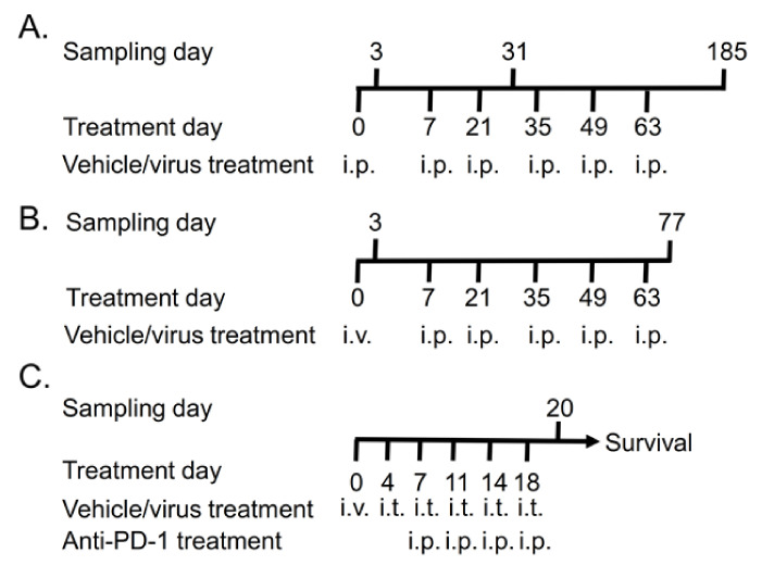
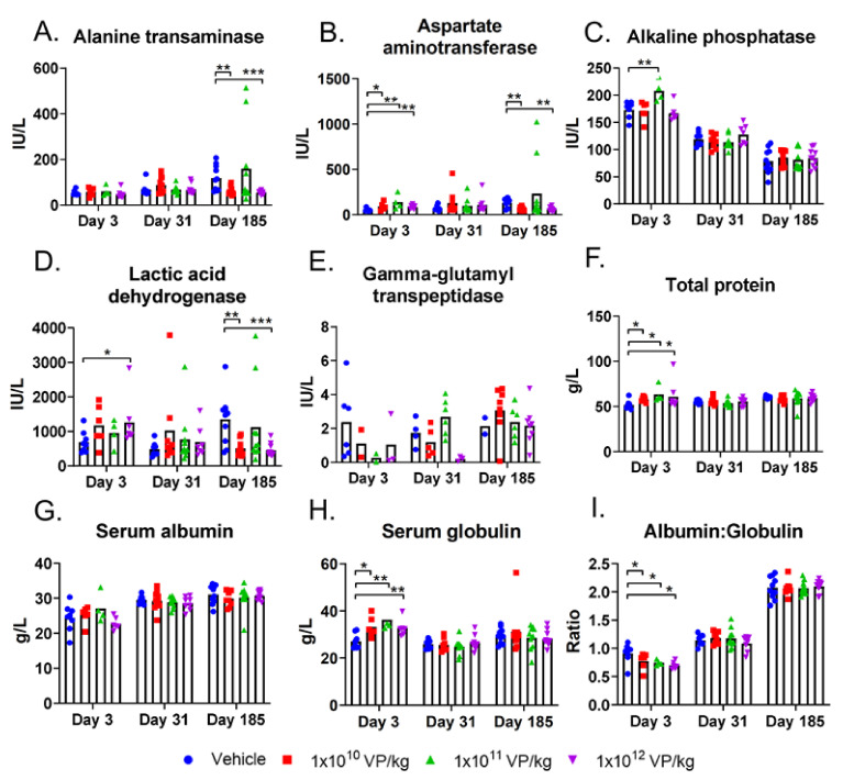
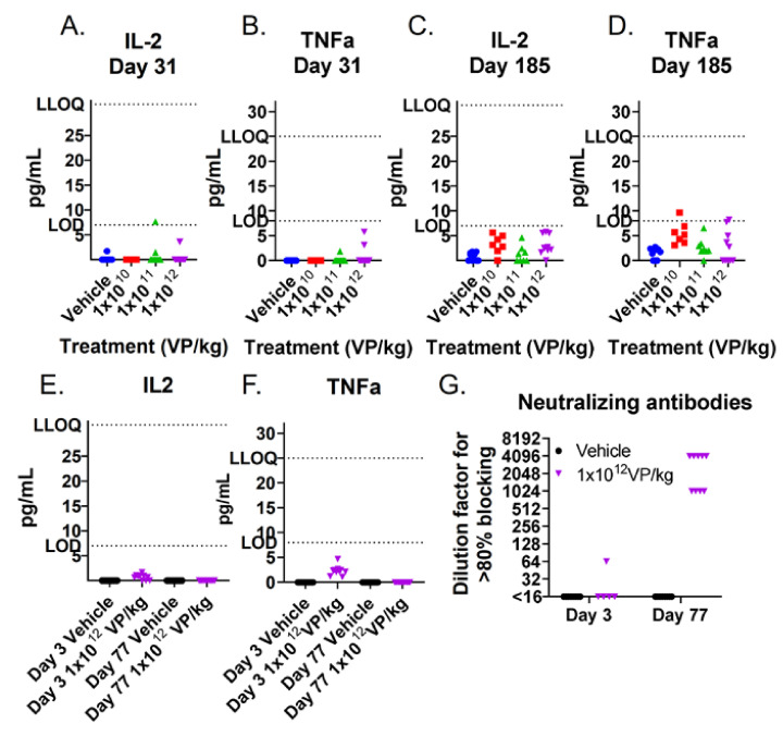
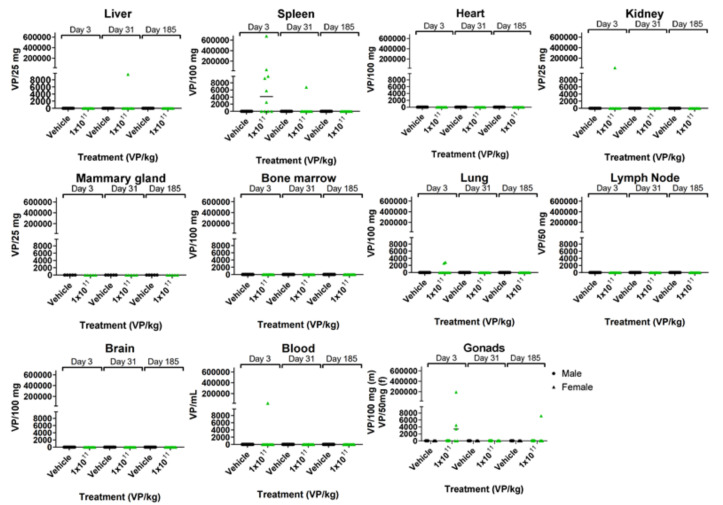
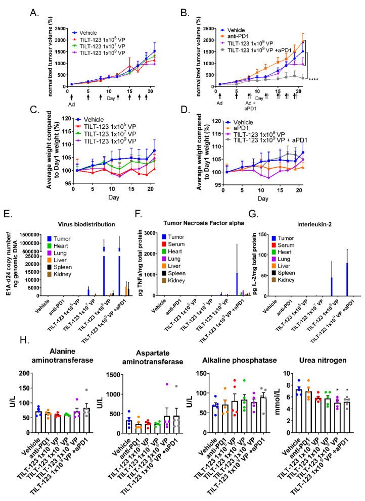
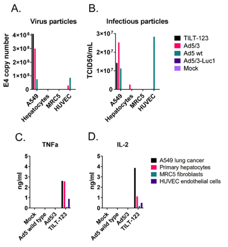
Oncolytic viruses provide a biologically multi-faceted treatment option for patients who cannot be cured with currently available treatment options. We constructed an oncolytic adenovirus, TILT-123, to support T-cell therapies and immune checkpoint inhibitors in solid tumors. Adenoviruses are immunogenic by nature, are easy to produce in large quantities, and can carry relatively large transgenes. They are the most commonly used gene therapy vectors and are well tolerated in patients. TILT-123 expresses two potent cytokines, tumor necrosis factor alpha and interleukin-2, to stimulate especially the T-cell compartment in the tumor microenvironment. Before entering clinical studies, the safety and biodistribution of TILT-123 was studied in Syrian hamsters and in mice. The results show that TILT-123 is safe in animals as monotherapy and in combination with an immune checkpoint inhibitor anti-PD-1. The virus treatment induces acute changes in circulating immune cell compartments, but the levels return to normal by the middle of the treatment period. The virus is rapidly cleared from healthy tissues, and it does not cause damage to vital organs. The results support the initiation of a phase 1 dose-escalation trial, where melanoma patients receiving a tumor-infiltrating lymphocyte therapy are treated with TILT-123 (NCT04217473).

Keywords: adenovirus; biodistribution; immunotherapy; oncolytic virus; safety.


Conflict of interest statement

R.H., R.K., and S.S. are employees of TILT Biotherapeutics Ltd. A.H., J.S., and V.C-C are employees and shareholders of TILT Biotherapeutics Ltd. A.H is a shareholder of Targovax ASA.

Figures

Figure 1
Figure 1
Treatment schedule for TILT-123 safety studies: (A) 15 male and female hamsters received saline or 1 × 10¹⁰, 1 × 10¹¹, or 1 × 10¹² virus particles (VP)/kg of TILT-123 six times intraperitoneally (i.p.). Five animals/sex/group were sacrificed 3, 31, and 185 days after the first treatment. (B) Ten male and female hamsters received saline, 1 × 10¹¹ VP/kg, or 1 × 10¹² VP/kg of TILT-123 once intravenously (i.v.) and five times i.p. Five animals/sex/group were sacrificed 3 days after the first injection and two weeks after the last injections (on day 77). (C) Human fibrosarcoma tumors were implanted subcutaneously to both flanks of nude NMRI mice. Nine days after implantation, the animals received 1 × 10⁵ VP, 1 × 10⁷ VP, or 1 × 10⁹ VP i.v. The following treatments were injected intratumorally (i.t.) Anti-PD-1 (100 µg) treatments were started on day 7 after two virus treatments. Five animals per group were sacrificed on day 20, and the rest (6–7 animals/group) continued in the survival study.
Figure 2
Figure 2
Circulating blood cell levels in hamsters: the hamsters received six TILT-123 injections i.p. at three different doses. Leucocytes (A), lymphocytes (B), large unstained cells (C), red blood cells (D), reticulocytes (E), thrombocytes (F), and myeloblast-derived cells (monocytes, basophils, eosinophils, and neutrophils) (G) were counted from the whole blood at three time points (3, 31, and 185 days after the first treatment). The data show the means plus standard error of mean. Statistical significance between the cell number in control group vs. treatment group on each time point was evaluated with the Mann–Whitney test (* p < 0.05).
Figure 3
Figure 3
Blood chemistry values in hamsters: the hamsters received six i.p. TILT-123 injections at three different doses. Alanine transaminase (A), aspartate aminotransferase (B), alkaline phosphatase (C), lactic acid dehydrogenase (D), gamma-glutamyl transpeptidase (E), total protein level (F), serum albumin (G), and serum globulin (H) were measured from hamster serum samples at three time points (3, 31, and 185 days after the first treatment). In addition, albumin to globulin ratio was calculated to depict the relative amounts of these proteins in serum (I). The data show means plus standard error of mean. Statistical significance between the molecule levels in the control group vs. treatment group on each time point was evaluated with the Mann–Whitney test (* p < 0.05; ** p < 0.01; *** p < 0.001).
Figure 4
Figure 4
TILT-123 transgene expression and neutralizing antibodies in hamster sera: human TNFa and IL-2 were analyzed from hamster sera by ELISA after three i.p. injections on day 31 (A,B) and after six injections at the end of the experiment (day 185) (C,D). In addition, the transgene expression (E,F) and Ad5/3 neutralizing antibodies (G) were measured after one i.v. injection (day 3) and two weeks after the last i.p. injection (day 77). LOD: Limit of Detection; LLOQ: Lower Limit of Quantification.
Figure 5
Figure 5
TILT-123 biodistribution in hamsters: the hamsters received six TILT-123 injections i.p. at the dose of 1 × 10¹¹ VP/kg. The presence of TILT-123 in hamster tissues was studied with qPCR at three time points (3, 31, and 185 days after the first treatment). Only values above the limit of quantification are shown.
Figure 6
Figure 6
Mice treated with TILT-123 and anti-PD-1 show tumor growth reduction. Nude mice bearing subcutaneous human fibrosarcoma tumors received TILT-123 (black arrows) and anti-PD-1 treatments (grey arrows) twice a week. Anti-PD-1 treatments started on day 7 and were given i.p. The first virus injection was given i.v., and the rest was given i.t. Tumor size was measured with a digital caliper (A,B), and animals were weighed (C,D) two to three times a week. Tumor size and animal weight at the beginning of the treatment period was set to 100%. Adenovirus genomes were detected from the tumors and healthy tissue samples (E) by qPCR and normalized against the genomic DNA amount or sample volume. TILT-123 transgenes human TNFa (F) and human IL-2 (G) were analyzed from tumors and healthy tissue samples with ELISA and were normalized to total protein contents in the samples. (H) Whole blood was collected from mice 48 h after last treatment (day 20). The serum was separated after blood coagulation. The data show mean plus standard error of mean. Statistical significance was evaluated with the log-linear mixed models analysis (tumor growth and weight development) or with one-way ANOVA. * p < 0.05; **** p < 0.0001; Ad = TILT-123 injection; aPD1 = anti-PD-1; VP = Virus Particles.
Figure 7
Figure 7
TILT-123 replicates selectively in cancer cells in vitro. A549 lung adenocarcinoma cells, human primary hepatocytes, MRC5 fibroblasts, and human vascular endothelial HUVEC cells were infected with TILT-123, Ad5/3 replicative control virus (similar selection device as TILT-123 but no cytokine payload), wild-type adenovirus type 5 (Ad5 wt), or replication-incompetent Ad5/3-Luc1 virus or left uninfected as a control (mock). (A) Virus genomes and (B) infectious virus particles were analyzed from the infected cells 72 h after infection. TILT-123 transgenes TNFa (C) and IL-2 (D) were analyzed from cell culture supernatants.

References

    1. Garber K. China approves world’s first oncolytic virus therapy for cancer treatment. J. Natl. Cancer Inst. 2006;98:298–300. doi: 10.1093/jnci/djj111. - DOI - PubMed
    1. Ranki T., Pesonen S., Hemminki A., Partanen K., Kairemo K., Alanko T., Lundin J., Linder N., Turkki R., Ristimaki A., et al. Phase I study with ONCOS-102 for the treatment of solid tumors-an evaluation of clinical response and exploratory analyses of immune markers. J. Immunother Cancer. 2016;4:1–18. doi: 10.1186/s40425-016-0121-5. - DOI - PMC - PubMed
    1. Lang F.F., Conrad C., Gomez-Manzano C., Yung W.K.A., Sawaya R., Weinberg J.S., Prabhu S.S., Rao G., Fuller G.N., Aldape K.D., et al. Phase I Study of DNX-2401 (Delta-24-RGD) Oncolytic Adenovirus: Replication and Immunotherapeutic Effects in Recurrent Malignant Glioma. J. Clin. Oncol. 2018 doi: 10.1200/JCO.2017.75.8219. - DOI - PMC - PubMed
    1. Nemunaitis J., Tong A.W., Nemunaitis M., Senzer N., Phadke A.P., Bedell C., Adams N., Zhang Y.A., Maples P.B., Chen S., et al. A phase I study of telomerase-specific replication competent oncolytic adenovirus (telomelysin) for various solid tumors. Mol. Ther. 2010;18:429–434. doi: 10.1038/mt.2009.262. - DOI - PMC - PubMed
    1. Eriksson E., Milenova I., Wenthe J., Stahle M., Leja-Jarblad J., Ullenhag G., Dimberg A., Moreno R., Alemany R., Loskog A. Shaping the Tumor Stroma and Sparking Immune Activation by CD40 and 4-1BB Signaling Induced by an Armed Oncolytic Virus. Clin. Cancer Res. 2017;23:5846–5857. doi: 10.1158/1078-0432.CCR-17-0285. - DOI - PubMed

Publication types

MeSH terms

Associated data