. 2021 Jan 27;13(2):96.
doi: 10.3390/toxins13020096.

Effective Locations for Injecting Botulinum Toxin into the Mentalis Muscle; Cadaveric and Ultrasonographic Study

Affiliations

Effective Locations for Injecting Botulinum Toxin into the Mentalis Muscle; Cadaveric and Ultrasonographic Study

Da-Yae Choi et al. Toxins (Basel). .

Abstract

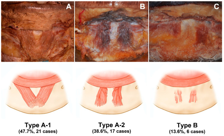
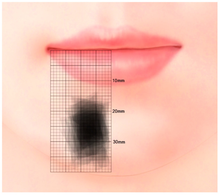
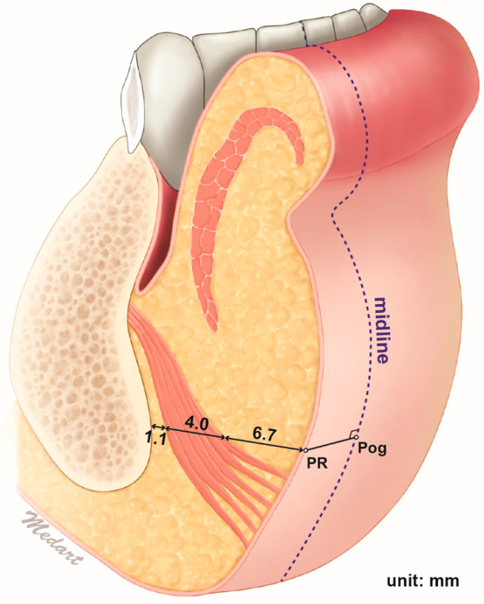
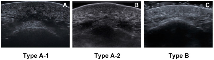
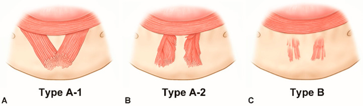
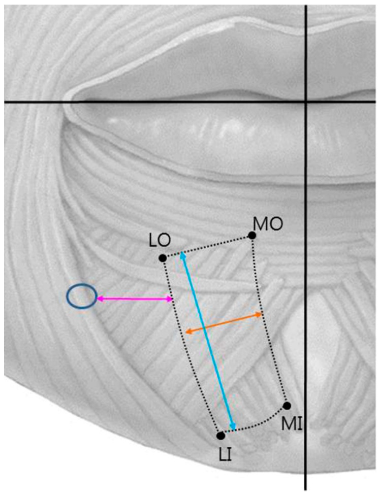
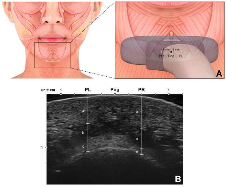
The mentalis muscle is now considered key structures when performing procedures for rejuvenating the lower face. The aim of this study was to determine the anatomical morphology and location of the mentalis muscle and thereby provide anatomical information for facilitating clinical procedures designed to rejuvenate the lower face. Forty-four adult hemifaces from five Thai cadavers and 21 Korean cadavers were dissected to identify the locations of the mentalis muscle. Sixty-six hemifaces from 33 healthy young Korean subjects were included in an ultrasonographic study. The depth of the mentalis muscle below the skin surface, the thickness of the mentalis muscle, and the distance from the bone to the mentalis muscle were measured at the two points that were 5 mm lateral to the most-prominent point of the chin. The mentalis muscle was classified into two types based to its shape: in type A (86.4%, 38 of the 44 cases) it was dome shaped in three dimensions, while in type B (13.6%, 6 of the 44 cases) it was flat. The mentalis muscle was present mostly at the area 5-10 mm from the midsagittal line and 20-30 mm from a horizontal line connecting the mouth corners. The mentalis muscle was present between depths of 6.7 to 10.7 mm below the skin. This new information about the location of the mentalis muscle may help when identifying the most effective and safe botulinum toxin injection points and depths during esthetic procedures for weakened facial rhytides on the lower face.

Keywords: botulinum toxin; facial rejuvenating; facial rhytides; injection procedure; mental crease; mentalis muscle.


Conflict of interest statement

The authors declare no conflict of interest. The funding sponsors had no role in the design of the study, in the collection, analyses, or interpretation of data; in the writing of the manuscript, and in the decision to publish the results.

Figures

Figure 1
Figure 1
Three types of mentalis muscle morphology. (A) Type A-1, two bilateral mentalis muscles merged with each other. (B) Type A-2, the two mentalis muscles were separated. (C) Type B, mentalis muscle was flat and comprised of only a few muscle fibers.
Figure 2
Figure 2
Distribution of the mentalis muscle relative to the facial midsagittal line and a horizontal line connecting the mouth corners. In most specimens the mentalis muscle was present at the area 5–10 mm from the facial midsagittal line and 20–30 mm from a horizontal line connecting the mouth corners.
Figure 3
Figure 3
Z-axis anatomy of the mentalis muscle. The depth of the mentalis muscles was measured at the point 5 mm left and right from the pogonion. The depth of the mentalis muscle below the skin surface, the thickness of the mentalis muscle, and the distance from the bone to the mentalis muscle were 6.7 ± 1.4, 4.0 ± 1.4, and 1.1 ± 1.0 mm, respectively. The mentalis muscle was present between mean depths of 6.7 to 10.7 mm below the skin surface.
Figure 4
Figure 4
Three types of mentalis muscle morphology observed in ultrasonographic images. B mode, transverse view, 15-MHz linear transducer. (A) In type A-1, the mentalis muscle is dome shaped, and the muscle fibers on each side are merged. (B) In type A-2, the mentalis muscle is dome shaped, but the muscle fibers on each side are separate. (C) In type B, the mentalis muscle was flat and comprised of thin fibers.
Figure 5
Figure 5
Images and illustrations demonstrating various morphologies of the mentalis muscle. Some of the muscle specimens were dome shaped, where the muscle fibers on each side are merged (A) or are separate (B). Other muscle specimens were flat and comprised of thin fibers (C).
Figure 6
Figure 6
Reference lines and measurement points. MO, medial origin point; LO, lateral origin point; MI, medial insertion point; LI, lateral insertion point; blue circle, mental foramen; yellow arrow, width of the mentalis muscle; blue arrow, length of the mentalis muscle; pink arrow, distance between the mental foramen and the lateral margin of the mentalis muscle.
Figure 7
Figure 7
Ultrasonographic analysis of the mentalis muscle. (A) Reference points for observing and measuring the thickness of the mentalis muscle. Pog, pogonion; PL, the point 5 mm left from the Pog; PR, the point 5 mm right from the Pog. A linear probe was applied horizontally at the Pog perpendicular to the skin surface to obtain the ultrasonographic image. (B) Ultrasonographic image and the parameters measured for the mentalis muscle and surrounding tissues (B mode, transverse view, 15-MHz linear transducer). Each parameter was measured at both PL and PR.; a, the depth of the mentalis muscle below the skin surface; b, the thickness of the mentalis muscle; c, the distance from the bone to the mentalis muscle were measured.

References

    1. Garfein E.S., Zide B.M. Chin ptosis: Classification, anatomy, and correction. Craniomaxillofacial Trauma Reconstr. 2008;1:1–14. doi: 10.1055/s-0028-1098968. - DOI - PMC - PubMed
    1. Hur M.-S., Kim H.-J., Choi B.-Y., Hu K.-S., Kim H.-J., Lee K.-S. Morphology of the mentalis muscle and its relationship with the orbicularis oris and incisivus labii inferioris muscles. J. Craniofacial Surg. 2013;24:602–604. doi: 10.1097/SCS.0b013e318267bcc5. - DOI - PubMed
    1. Zide B.M., McCarthy J. The mentalis muscle: An essential component of chin and lower lip position. Plast. Reconstr. Surg. 1989;83:413–420. doi: 10.1097/00006534-198903000-00001. - DOI - PubMed
    1. Kim H.-J., Seo K.K., Lee H.-K., Kim J. Clinical Anatomy of the Face for Filler and Botulinum Toxin Injection. Springer; Singapore: 2016. General Anatomy of the Face and Neck; pp. 1–53.
    1. Coleman K.R., Carruthers J. Combination therapy with BOTOX™ and fillers: The new rejuvnation paradigm. Dermatol. Ther. 2006;19:177–188. doi: 10.1111/j.1529-8019.2006.00072.x. - DOI - PubMed

Publication types

Substances

LinkOut - more resources