. 2020 Sep 22;15(1):100.
doi: 10.1186/s13020-020-00381-3.

Micronized sacchachitin promotes satellite cell proliferation through TAK1-JNK-AP-1 signaling pathway predominantly by TLR2 activation

Affiliations

Micronized sacchachitin promotes satellite cell proliferation through TAK1-JNK-AP-1 signaling pathway predominantly by TLR2 activation

Meng-Huang Wu et al. Chin Med. .

Erratum in

Abstract

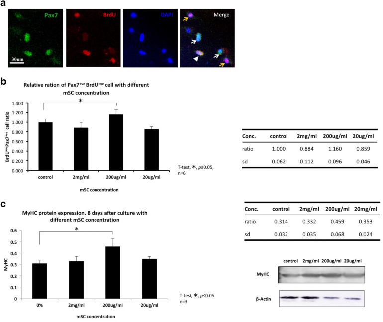
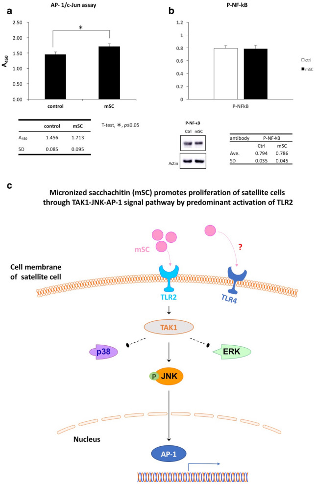
Background: Ganoderma sp., such as Ganoderma tsugae (GT), play an important role in traditional Chinese medicine. Ganoderma sp. contains several constituents, including Sacacchin, which has recently drawn attention because it can not only enhance the repair of muscle damage but also strengthen the muscle enforcement. Although Ganoderma sp. have a therapeutic effect for neuromuscular disorders, the underlying mechanism remains unclear. This study investigated the effect and underlying molecular mechanism of micronized sacchachitin (mSC) on satellite cells (SCs), which are known as the muscle stem cells.

Methods: The myogenic cells, included SCs (Pax7+) were isolated from tibialis anterior muscles of a healthy rat and were cultured in growth media with different mSC concentrations. For the evaluation of SC proliferation, these cultivated cells were immunostained with Pax7 and bromodeoxyuridine assessed simultaneously. The molecular signal pathway was further investigated by using Western blotting and signal pathway inhibitors.

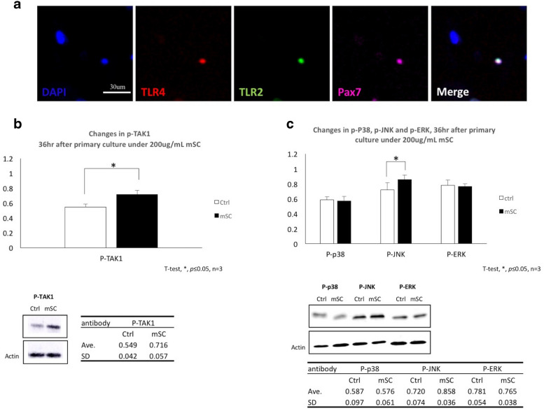
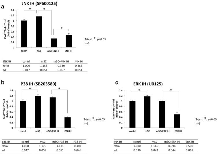
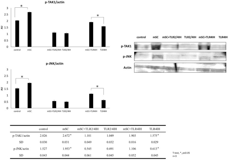
Results: Our data revealed that 200 µg/mL mSC had an optimal capability to significantly enhance the SC proliferation. Furthermore, this enhancement of SC proliferation was verified to be involved with activation of TAK1-JNK-AP-1 signaling pathway through TLR2, whose expression on SC surface was confirmed for the first time here.

Conclusion: Micronized sacchachitin extracted from GT was capable of promoting the proliferation of SC under a correct concentration.

Keywords: MAPK signal pathway; Muscle regeneration; Sacchachitin; Satellite cells; TAK1-JNK-AP-1 signaling pathway.


Conflict of interest statement

The authors report no conflicts of interests in this work.

Figures

Fig. 1
Fig. 1
Micronized sacchachitin (mSC) in concentration of 200ug/mL significantly enhanced the proliferation of Pax7 + ve SCs isolated from TA muscle. a Co-immunostaining using anti-Pax7 antibody and BrdU assay was performed 2 days after cell plating. Pax7 single positive (Pax7 + ve, white arrow), BrdU single positive (BrdU + ve, yellow arrow) and Pax7-BrdU double positive (Pax7 + ve BrdU + ve, white arrow head) were counted within each group. Scale bar is 30 μm. b Micronized sacchachitin (mSC) of 200 μg/ml significantly enhanced the proliferation of Pax7 + ve cells (relative to control, 1.160 ± 0.96, *p < 0.05, n = 6, one-way ANOVA with Dunnett’s multiple comparisons). c 8 days after cell plating, MyHC protein within mediums contained different mSC concentration was evaluated through Western blotting assay. Compatible with the results of Pax7 + ve BrdU + ve cell number, mSC of 200 μg/ml showed the highest MyHC (Myosin Heavy Chain) protein expression (0.459 ± 0.068, *p < 0.05, one-way ANOVA with Dunnett’s multiple comparisons)
Fig. 2
Fig. 2
Toll-like receptor 2 and TLR4 were expressed upon SC and the signals of its downstream, TAK1- JNK but not p38, ERK was activated by mSC. a The presence of TLR2/4 upon Pax7 + ve SCs were confirmed through immunostaining. Scale bar is 30 μm. b Relative to control, a significant elevation of pTAK-1 was admitted under mSC 200 μg/mL (0.716 ± 0.057, *p < 0.05, n = 3, unpaired t-test). c Significant elevation was only observed with p-JNK but p-p38 nor p-ERK (0.858 ± 0.036, p < 0.05, 0.576 ± 0.061, p = 0.258, 0.765 ± 0.038, p = 0.384, n = 3, unpaired t-test)
Fig. 3
Fig. 3
The proliferation of SC induced by mSC could be effectively suppressed by JNK-inhibitor but p38-inhibitor nor ERK-inhibitor. a The effective blockage of the enhanced SC proliferation under mSC 200 μg/ml was only observed with JNK inhibitor (mSC + IH relative to JNK IH SP600125, 0.330 ± 0.057 and 0.463 ± 0.054 respectively, p = 0.0570, n = 3), b but not with p38 inhibitor (mSC + IH relative to p38 IH SB203580, 1.131 ± 0.051 and 0.389 ± 0.046 respectively, ****p < 0.0001, n = 3) nor c ERK inhibitor (mSC + IH relative to ERK IH U0126, 0.994 ± 0.044 and 0.500 ± 0.068, ****p < 0.0001, respectively, n = 3, one-way ANOVA with Tukey’s multiple comparisons)
Fig. 4
Fig. 4
Micronized sacchachitin (mSC) induced JNK/MAPK phosphorylation was predominantly initiated by the activation of TLR2 and mediated by TAK1. The protein level of TAK1 and JNK/MAPK were significantly suppressed by TLR2/4-inhibitor (TLR2/4 IH, 1.10 ± 0.05, p = 0.07, 0.55 ± 0.06, p = 0.13, n = 3, unpaired t-test) but not by TLR4-inhibitor (TLR4IH, 1.903 ± 0.016, 1.106 ± 0.052, *p < 0.05, n = 3, unpaired t-test)
Fig. 5
Fig. 5
AP-1 transcriptional activity was significantly enhanced by mSC. a The AP-1 transcriptional activity of the TA muscle isolated myogenic cells, included Pax7 + ve SCs, which were cultivated with mSC (200 μg/mL) contained medium was significantly enhanced relative to that of the control (1.713 ± 0.095, *p < 0.05, n = 3, unpaired t-test). b There was no remarkable increased in phosphorylation of NF-κB admitted (0.786 ± 0.045, p = 0.481, n = 3, unpaired t-test). c The enhancement of SC proliferation induced by mSC was predominantly through TLR2 and the activation of TAK 1-JNK-AP-1 signal pathway. Interestingly, JNK was solely activated among the MAPK signaling

References

    1. Pang X, Chen Z, Gao X, Liu W, Slavin M, Yao W, Yu LL. Potential of a novel polysaccharide preparation (GLPP) from Anhui-grown Ganoderma lucidum in tumor treatment and immunostimulation. J Food Sci. 2007;72(6):S435–S442. - PubMed
    1. Su CH, Sun CS, Juan SW, Hu CH, Ke WT, Sheu MT. Fungal mycelia as the source of chitin and polysaccharides and their applications as skin substitutes. Biomaterials. 1997;18(17):1169–1174. - PubMed
    1. Chien RC, Yen MT, Tseng YH, Mau JL. Chemical characteristics and anti-proliferation activities of Ganoderma tsugae polysaccharides. Carbohydr Polym. 2015;128:90–98. - PubMed
    1. Chen ML, Hsieh CC, Chiang BL, Lin BF. Triterpenoids and polysaccharide fractions of Ganoderma tsugae exert different effects on antiallergic activities. Evid Based Complement Alternat Med. 2015;2015:754836. - PMC - PubMed
    1. Zjawiony JK. Biologically active compounds from Aphyllophorales (polypore) fungi. J Nat Prod. 2004;67(2):300–310. - PubMed