. 2021 Jan 29;14(1):21.
doi: 10.1186/s13045-020-01024-8.

Targeting B7-H3 via chimeric antigen receptor T cells and bispecific killer cell engagers augments antitumor response of cytotoxic lymphocytes

Affiliations

Targeting B7-H3 via chimeric antigen receptor T cells and bispecific killer cell engagers augments antitumor response of cytotoxic lymphocytes

Jie Liu et al. J Hematol Oncol. .

Abstract

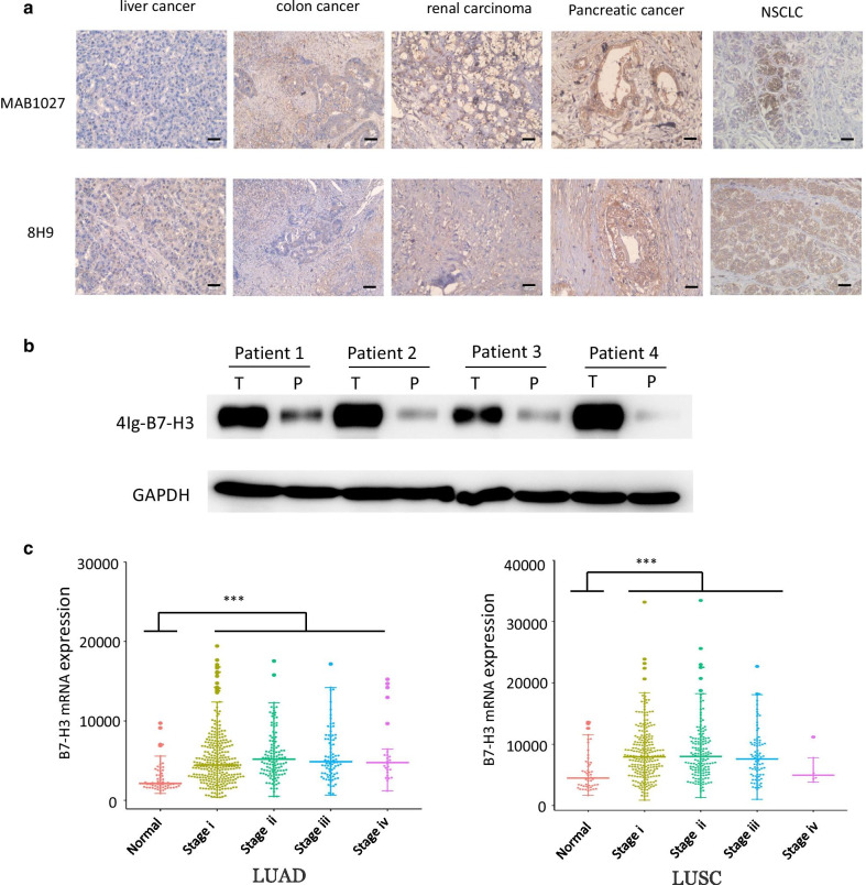
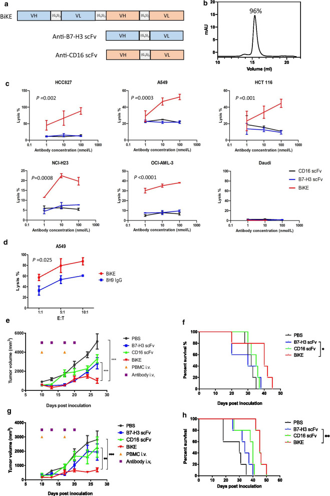
Background: B7-H3, an immune-checkpoint molecule and a transmembrane protein, is overexpressed in non-small cell lung cancer (NSCLC), making it an attractive therapeutic target. Here, we aimed to systematically evaluate the value of B7-H3 as a target in NSCLC via T cells expressing B7-H3-specific chimeric antigen receptors (CARs) and bispecific killer cell engager (BiKE)-redirected natural killer (NK) cells.

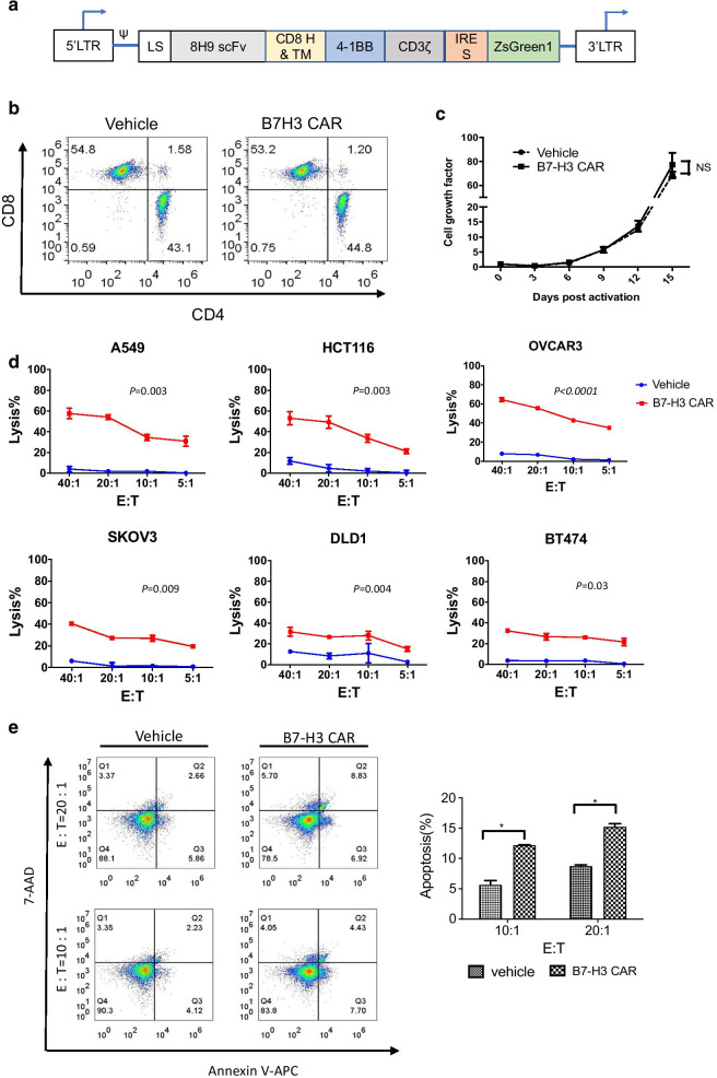
Methods: We generated B7-H3 CAR and B7-H3/CD16 BiKE derived from an anti-B7-H3 antibody omburtamab that has been shown to preferentially bind tumor tissues and has been safely used in humans in early-phase clinical trials. Antitumor efficacy and induced-immune response of CAR and BiKE were evaluated in vitro and in vivo. The effects of B7-H3 on aerobic glycolysis in NSCLC cells were further investigated.

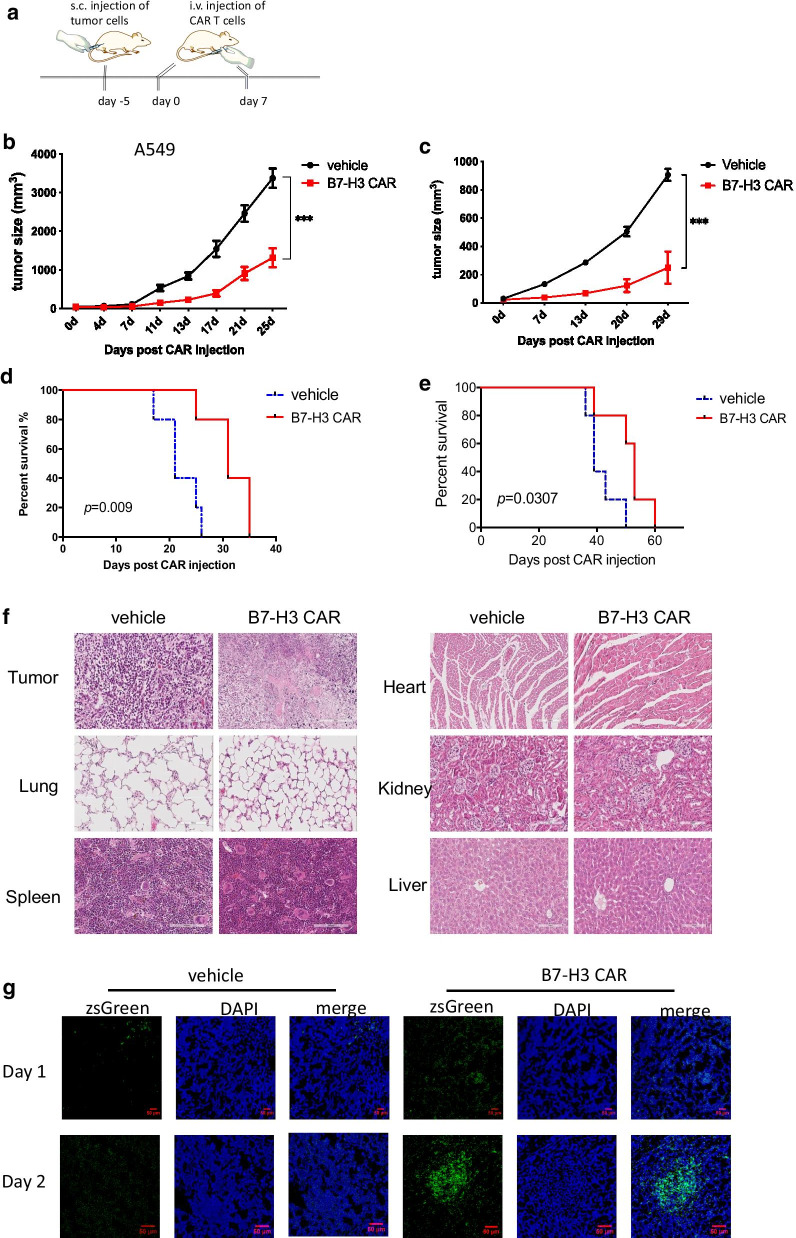
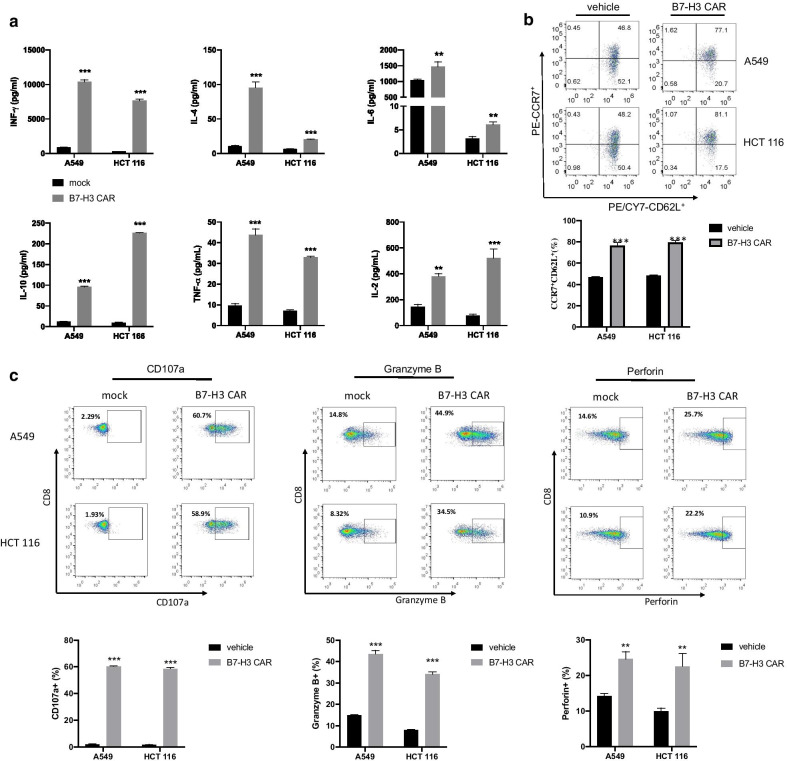
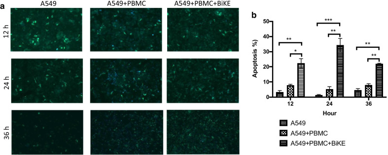
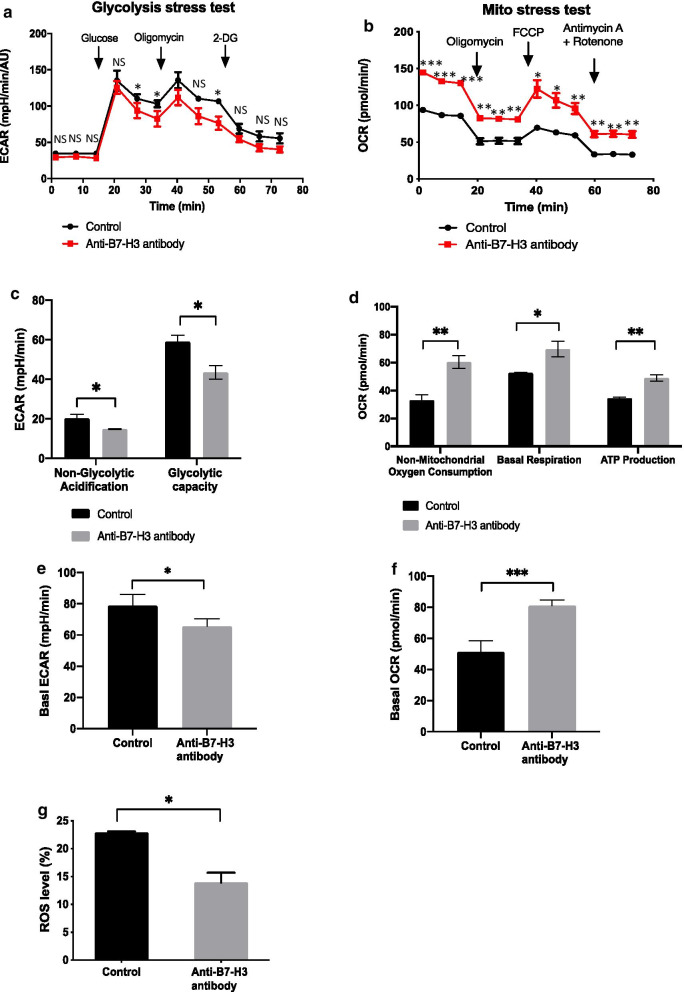
Results: B7-H3 CAR-T cells effectively inhibited NSCLC tumorigenesis in vitro and in vivo. B7-H3 redirection promoted highly specific T-cell infiltration into tumors. Additionally, NK cell activity could be specially triggered by B7-H3/CD16 BiKE through direct CD16 signaling, resulting in significant increase in NK cell activation and target cell death. BiKE improved antitumor efficacy mediated by NK cells in vitro and in vivo, regardless of the cell surface target antigen density on tumor tissues. Furthermore, we found that anti-B7-H3 blockade might alter tumor glucose metabolism via the reactive oxygen species-mediated pathway.

Conclusions: Together, our results suggest that B7-H3 may serve as a target for NSCLC therapy and support the further development of two therapeutic agents in the preclinical and clinical studies.

Keywords: B7-H3; BiKE; Bispecific antibody; CAR T; Chimeric antigen receptor; Non-small cell lung cancer; PD-L1.


Conflict of interest statement

The authors declare that they have no conflicts of interest.

Figures

Fig. 1
Fig. 1
Expression of B7-H3 in human tissues. a IHC of FFPE tissues was used to evaluate B7-H3 expression in various human tumors. The sale bar represents 20 μm. b Western blot analysis of B7-H3 in tumor (T) and paracancerous (P) tissues from four NSCLC patients. The tissue lysates were probed with the goat anti-B7-H3 antibody. The levels of GAPDH were utilized as loading control. c Differential expression of B7-H3 in normal lung tissues, lung adenocarcinoma (LUAD) and squamous cell carcinoma (LUSC) in different stages was analyzed using RNA-seq datasets from TCGA. Significant differences were calculated using Student’ t test (*p < 0.05, **p < 0.01, ***p < 0.001)
Fig. 2
Fig. 2
Generation of B7-H3 CAR- T cells and their in vitro antitumor activity. a Schematic diagram of the CAR construct. CAR gene was cloned into a lentiviral vector, which contained an internal ribosome entry site (IRES)-green fluorescence protein (ZsGreen) sequence. The resulting CAR was composed of the leader sequence (LS), a scFv, a hinge (H) region, a CD8α transmembrane domain (TM), along with the 4-1BB domain and the CD3ζ chain. b Subsets of B7-H3 CAR-T cells were derived from a single healthy donor. T cells were stained with anti-CD3, anti-CD4, or anti-CD8 antibodies and then analyzed using flow cytometry. c Proliferation of B7-H3 CAR-T cells and vehicle T cells activated by anti-CD3/CD28 beads. d Cytotoxicity of B7-H3 CAR-T cells (red line) and vehicle T cells (blue line) cells against various tumor cell lines. e Apoptosis in target A549 cells induced by B7-H3 CAR-T cells and vehicle T cells as control at different E:T ratios. Representative graphs are shown. The p values of the difference between the CAR group and the control group were analyzed using ANOVA
Fig. 3
Fig. 3
Therapeutic effects of B7-H3 CAR-T cells in tumor xenografts. a Treatment scheme of B7-H3 CAR-T cells in A549 or HCT 116 xenograft models. be Tumor growth in A549-injected mice b or HCT 116-injected mice c was measured using digital caliper, and tumor area was calculated (n = 5). Mean values per treatment group are shown. The p values of treatment were analyzed using ANOVA. Survival curves of A549-injected mice d or HCT 116-injected mice e are shown. The p values of survival curves were analyzed using the log-rank (Mantel–Cox) test. Data are presented as the mean ± standard deviation. *p < 0.05, **p < 0.01, ***p < 0.001. f H&E staining of major organs in mice treated with CAR-T cells. g  Tumor infiltration of B7-H3 CAR-T cells in A549-injected mice. On days 1 and 2, the tumors were harvested, sliced, fixed and imaged in the fluorescence microscope. Representative images are shown. ZsGreen is used as the reporter of CAR-T cells. Representative images are shown
Fig. 4
Fig. 4
Functional analysis of B7-H3 CAR-T cells. a Cytokine production in B7-H3 CAR and vehicle T cells. Effector T cells were incubated with target A549 or HCT 116 cells at a ratio of 10:1 for 24 h. The concentrations of cytokines were measured using flow cytometry. b Memory cell phenotypes of B7-H3 CAR-T cells. Effector T cells were incubated with target A549 or HCT 116 cells at a ratio of 10:1 for 24 h. Expression of CD62L and CCR7 on T cells was identified with staining with anti-CD62L and anti-CCR7 antibodies, respectively, and measured using flow cytometry. c Degranulation markers in B7-H3 CAR T cells. Effector T cells were incubated with target A549 or HCT 116 cells at a ratio of 10:1 for 6 h (CD107a) or 24 h (granzyme B and perforin). Expressions of CD107a, intracellular granzyme B and perforin staining on T cells were stained with anti CD107a, granzyme B and perforin antibodies and measured by flow cytometry. Data from 2–3 independent experiments are presented as the mean ± standard deviation. *p < 0.05, **p < 0.01, ***p < 0.001. The p values were analyzed using a Student’ t test
Fig. 5
Fig. 5
B7-H3/CD16 BiKE induced antitumor effect mediated by NK cells in vitro and in vivo. a Construct diagrams of B7-H3/CD16 BiKE, anti-B7-H3 scFv, and anti-CD16 scFv. b Purity of B7-H3/CD16 BiKE determined by size-exclusion chromatography. c Cytotoxicity of B7-H3/CD16 BiKE (red line), anti-B7-H3 scFv (blue line) and anti-CD16 scFv (black line) against different tumor cell lines by PBMC as effectors. d Comparison of ADCC induced by B7-H3/CD16 BiKE and anti-B7-H3 IgG 8H9. PBMCs were used as effectors and A549 cells were used as targets. Representative graphs are shown. For the experiments cd, the p values of the difference between BiKE and control groups were analyzed using ANOVA. eh Therapeutic effects of B7-H3/CD16 BiKE in A549 and NCI-H23 xenografted mice. Antibodies were administered (100 μg per dose) intravenously twice a week for a total of four doses. Human PBMCs (10 million per iv injection) were delivered intravenously twice. Tumor growth in A549-injected mice e or HCT 116-injected mice g was measured using digital caliper, and tumor area was calculated (n = 5). Mean values per treatment group are shown.  Survival curves of A549-injected mice f or HCT 116-injected mice (h) are shown. The p values of treatment were analyzed using ANOVA. The p values of survival curves were analyzed using the log-rank (Mantel–Cox) test. 
Fig. 6
Fig. 6
Dynamic caspase-3 activation in apoptotic cells in response to B7-H3/CD16 BiKE. a FRET imaging of A549-C3 cells live cells appeared in green color and apoptotic cells in blue color. A549-C3 cells were treated with B7-H3/CD16 BiKE (5 μg/mL) with PBMCs at E:T = 10 for 12,24, and 36 h. b Statistical analysis of apoptotic rate at different time points. Representative images are shown. Sample size: 1 × 105 target cells. The p values were analyzed using a Student’ t test
Fig. 7
Fig. 7
Anti-B7-H3 blockade influences on glucose metabolism and intracellular ROS in A549 tumor cells. af Extracellular acidification rate and oxygen consumption rate measurements using the seahorse extracellular flux analyzer. After A549 cells were treated with either anti-B7-H3 or control antibodies for 24 h, ECAR and OCAR were examined using the Mito Stress Cell and Glycolysis Stress assays. ECAR (a, e) and OCR (b, f) metabolic profiles are shown. c Nonglycolytic acidification and glycolytic capacity were derived from Glycolysis Stress assay results. d Nonmitochondrial respiration, ATP production and basal respiration were derived from Mito Stress Cell results. g Intracellular ROS measurement in tumor cells. A549 cells were labeled with 2,7-dichlorofluorescin diacetate as a probe and treated with either anti-B7-H3 or control antibodies. Intracellular ROS signals were measured using flow cytometry. Data from two independent experiments are presented as the mean ± standard deviation. (*p < 0.05, **p < 0.01, ***p < 0.001). The p values were analyzed using a Student’ t test

Similar articles

Cited by

References

    1. Oser MG, Niederst MJ, Sequist LV, Engelman JA. Transformation from non-small-cell lung cancer to small-cell lung cancer: molecular drivers and cells of origin. Lancet Oncol. 2015;16:e165–e172. doi: 10.1016/S1470-2045(14)71180-5. - DOI - PMC - PubMed
    1. Riihimaki M, Hemminki A, Fallah M, Thomsen H, Sundquist K, Sundquist J, et al. Metastatic sites and survival in lung cancer. Lung Cancer. 2014;86:78–84. doi: 10.1016/j.lungcan.2014.07.020. - DOI - PubMed
    1. Popper HH. Progression and metastasis of lung cancer. Cancer Metastasis Rev. 2016;35:75–91. doi: 10.1007/s10555-016-9618-0. - DOI - PMC - PubMed
    1. Zappa C, Mousa SA. Non-small cell lung cancer: current treatment and future advances. Transl Lung Cancer Res. 2016;5:288–300. doi: 10.21037/tlcr.2016.06.07. - DOI - PMC - PubMed
    1. Chen R, Manochakian R, James L, Azzouqa AG, Shi H, Zhang Y, et al. Emerging therapeutic agents for advanced non-small cell lung cancer. J Hematol Oncol. 2020;13:58. doi: 10.1186/s13045-020-00881-7. - DOI - PMC - PubMed

Publication types

MeSH terms