. 2021 Jul;35(7):1869-1878.
doi: 10.1038/s41433-021-01408-1. Epub 2021 Jan 29.

Automated MRI-based quantification of posterior ocular globe flattening and recovery after long-duration spaceflight

Affiliations

Automated MRI-based quantification of posterior ocular globe flattening and recovery after long-duration spaceflight

Stuart H Sater et al. Eye (Lond). 2021 Jul.

Abstract

Background/objectives: Spaceflight associated neuro-ocular syndrome (SANS), a health risk related to long-duration spaceflight, is hypothesized to result from a headward fluid shift that occurs with the loss of hydrostatic pressure gradients in weightlessness. Shifts in the vascular and cerebrospinal fluid compartments alter the mechanical forces at the posterior eye and lead to flattening of the posterior ocular globe. The goal of the present study was to develop a method to quantify globe flattening observed by magnetic resonance imaging after spaceflight.

Subjects/methods: Volumetric displacement of the posterior globe was quantified in 10 astronauts at 5 time points after spaceflight missions of ~6 months.

Results: Mean globe volumetric displacement was 9.88 mm3 (95% CI 4.56-15.19 mm3, p < 0.001) on the first day of assessment after the mission (R[return]+ 1 day); 9.00 mm3 (95% CI 3.73-14.27 mm3, p = 0.001) at R + 30 days; 6.53 mm3 (95% CI 1.24-11.83 mm3, p < 0.05) at R + 90 days; 4.45 mm3 (95% CI -0.96 to 9.86 mm3, p = 0.12) at R + 180 days; and 7.21 mm3 (95% CI 1.82-12.60 mm3, p < 0.01) at R + 360 days.

Conclusions: There was a consistent inward displacement of the globe at the optic nerve, which had only partially resolved 1 year after landing. More pronounced globe flattening has been observed in previous studies of astronauts; however, those observations lacked quantitative measures and were subjective in nature. The novel automated method described here allows for detailed quantification of structural changes in the posterior globe that may lead to an improved understanding of SANS.


Conflict of interest statement

BAM has received grant support from Genentech, Minnetronix Neuro, Biogen, Voyager Therapeutics, and Alcyone Lifesciences. BAM is vice president of Research, Precision Delivery, & CSF Sciences at Alcyone Therapeutics. SHS is an employee at alcyone therapeutics. BAM is scientific advisory board member for the Chiari and Syringomyelia Foundation and serves as a consultant to SwanBio Therapeutics, Cerebral Therapeutics, Minnetronix Neuro, Genentech and CereVasc.

Figures

Fig. 1
Fig. 1. Methods for segmenting T2-weighted axial ocular MRI.
A MR images were radially resliced, and B automatically segmented. C Zoomed in view of the posterior globe (green square on B), showing the sclera (hypointense region), the putative retinal/choroidal complex, and the boundary identified by our segmentation scheme (red curve). D 3D reconstructed downsampled point cloud, and E the registration of point clouds. F Example of pre- and postflight distance maps and the resulting differential displacement map for the posterior surface of one eye with notable globe flattening, within the 4 mm region of interest (blue circle) around the optic nerve head on the displacement map (colour figure online).
Fig. 2
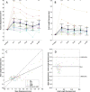
Fig. 2. Plots showing volume displacement and ocular biometry changes after spaceflight.
Plots showing pre- to postflight A MRI-assessed volume changes in the posterior ocular globe and B optical biometry-assessed axial length decreases at multiple time points after return to earth (R+; days) postflight. All changes are referenced to preflight values. Triangles and squares represent right and left eyes, respectively. Each subject is shown in a different colour, with yellow representing the subject diagnosed with grade 1 optic disc oedema. Black markers and error bars represent mean values with 95% confidence intervals and black stars indicate statistical significance from preflight baseline. Volume displacement within a 4 mm radius of the optic nerve head was averaged and compared with ocular axial length decreases (pre-post) as measured by ocular biometry using C correlation and D Bland–Altman plots. Note: B was created using data adapted from Macias et al. [5] *p < 0.05, **p < 0.01, ***p < 0.001 (colour figure online).
Fig. 3
Fig. 3. Summary of all globe displacement maps for each subject (within a 4 mm radius around the optic nerve head) at multiple time points (R + 1, R + 30, R + 90, R + 180, and R + 360 days).
OS and OD refer to left and right eye respectively. Grey boxes indicate that data were not available for that time point. The subject with the most severe displacement (subject 2, OD) was clinically diagnosed with grade 1 optic disc oedema via fundus imaging.

References

    1. Kramer LA, Sargsyan AE, Hasan KM, Polk JD, Hamilton DR. Orbital and intracranial effects of microgravity: findings at 3-T MR imaging. Radiology. 2012;263:819–27. doi: 10.1148/radiol.12111986. - DOI - PubMed
    1. Lee AG, Mader TH, Gibson CR, Brunstetter TJ, Tarver WJ. Space flight-associated neuro-ocular syndrome (SANS) Eye. 2018;32:1164–7. doi: 10.1038/s41433-018-0070-y. - DOI - PMC - PubMed
    1. Mader TH, Gibson CR, Otto CA, Sargsyan AE, Miller NR, Subramanian PS, et al. Persistent asymmetric optic disc swelling after long-duration space flight: implications for pathogenesis. J Neuroophthalmol. 2017;37:133–9. doi: 10.1097/WNO.0000000000000467. - DOI - PubMed
    1. Mader TH, Gibson CR, Pass AF, Kramer LA, Lee AG, Fogarty J, et al. Optic disc edema, globe flattening, choroidal folds, and hyperopic shifts observed in astronauts after long-duration space flight. Ophthalmology. 2011;118:2058–69. doi: 10.1016/j.ophtha.2011.06.021. - DOI - PubMed
    1. Macias BR, Patel NB, Gibson CR, Samuels BC, Laurie SS, Otto C, et al. Association of long-duration spaceflight with anterior and posterior ocular structure changes in astronauts and their recovery. JAMA Ophthalmol. 2020;138:553–9. doi: 10.1001/jamaophthalmol.2020.0673. - DOI - PMC - PubMed

LinkOut - more resources