. 2021 Mar;226(2):575-600.
doi: 10.1007/s00429-020-02201-8. Epub 2021 Jan 30.

Development of the mouse anterior amygdalar radial unit marked by Lhx9-expression

Affiliations

Development of the mouse anterior amygdalar radial unit marked by Lhx9-expression

Elena Garcia-Calero et al. Brain Struct Funct. 2021 Mar.

Abstract

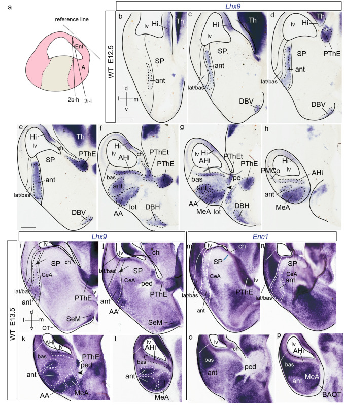
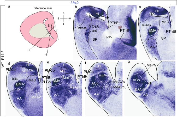
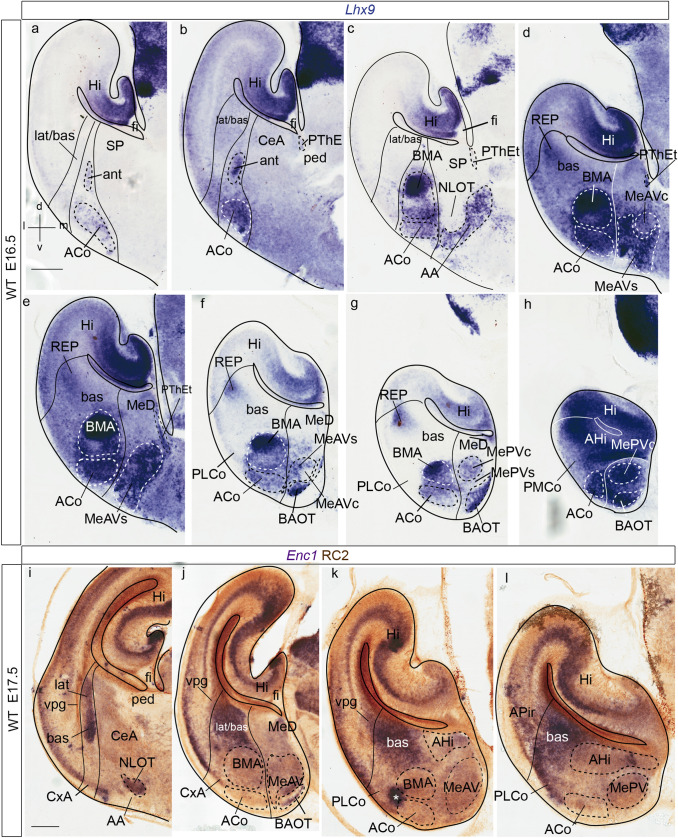
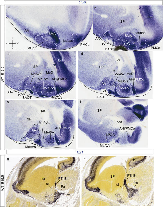
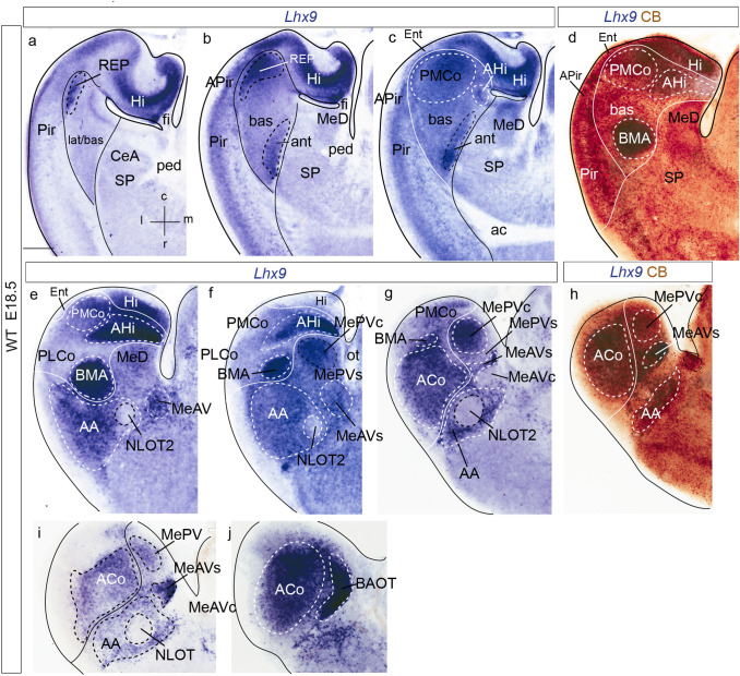
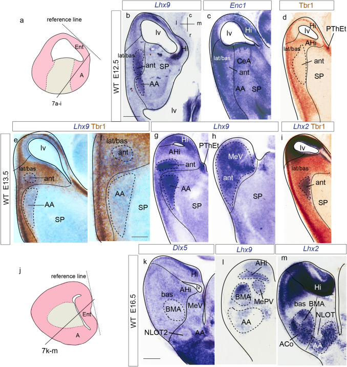
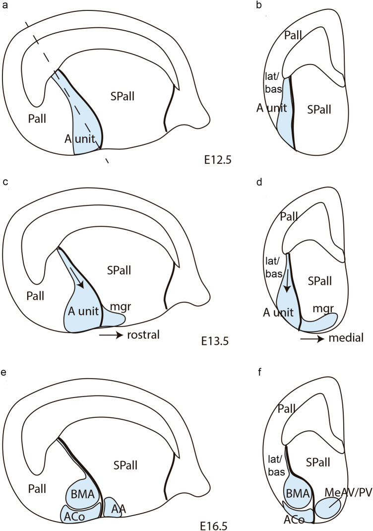
The amygdala in mammals plays a key role in emotional processing and learning, being subdivided in pallial and subpallial derivatives. Recently, the cortical ring model and the pallial amygdalar radial model (Puelles et al. 2019; Garcia-Calero et al. 2020) described the pallial amygdala as an histogenetic field external to the allocortical ring, and subdivided it in five major radial domains called lateral, basal, anterior, posterior and retroendopiriform units. The anterior radial unit, whose cells typically express the Lhx9 gene (see molecular profile in Garcia-Calero et al. 2020), is located next to the pallial/subpallial boundary. This radial domain shows massive radial translocation and accumulation of its derivatives into its intermediate and superficial strata, with only a glial palisade representing its final periventricular domain. To better understand the development of this singular radial domain, not described previously, we followed the expression of Lhx9 during mouse amygdalar development in the context of the postulated radial subdivisions of the pallial amygdala and other telencephalic developmental features.

Keywords: Medial amygdala; Pallial amygdala; Pallio-subpallial boundary; Pallium; Radial amygdalar model; Ventral pallium.


Conflict of interest statement

The authors declare that they have no conflict of interest.

Figures

Fig. 1
Fig. 1
Amygdalar Lhx9 expression in stage E12.5 mouse embryos. a, i Schematic representation of section planes: a amygdalar radial plane for figures (bh); i oblique sagittal plane for figures (jo). bh Lhx9 expression images ordered from rostral to caudal levels; b orientation in upper left-hand corner. The limits between the lateral/basal complex and the olfactory cortex, as well as between the lateral/basal complex and the anterior radial unit, or the latter and subpallium are indicated with thin black lines. jo Sagittal Lhx9 expression images ordered from lateral to medial levels; j orientation in upper right-hand corner. For abbreviations, see list. Scale bars in bo represent 300 µm
Fig. 2
Fig. 2
Lhx9 and Enc1 expression in mouse amygdalar region at embryonic stages E12.5 and E13.5. a Schematic representation of section plane. bh Lhx9 expression at stage E12.5 ordered from rostral to caudal levels; b orientations indicated at the bottom left-hand corner. il Lhx9 expression at stage E13.5 in a slightly different amygdalar radial plane (see a), ordered from rostral to caudal levels. mp Enc1 expression at stage E13.5, ordered from rostral to caudal levels, in alternate sections from the same embryo as in (il); i, j black arrow indicates a Lhx9-negative gap in the deep strata of the anterior amygdalar radial unit; orientation indicated in the bottom left-hand corner of (i). g, k black arrowhead point to positive Lhx9 cells in the medial amygdalar surface with possible source in PThEt. The limits between the lateral/basal complex and olfactory cortex, between the lateral/basal complex and the anterior radial unit, or the latter and subpallium are indicated with black or white lines. For abbreviations, see list. Scale bars represent 300 µm (bh) and 350 µm (ip)
Fig. 3
Fig. 3
Lhx9 expression in mouse amygdalar region at embryonic stage E14.5, in nearly horizontal sections ordered from dorsal to ventral levels. Note lack of continuity of anterior unit signal with the ventricle. a Schematic representation of section plane for Figs.3 and 4; b orientation indicated in the upper right-hand corner. The limits between the posterior radial unit and the hippocampus, the lateral/basal complex and the cortex, as well as between the lateral/basal complex and the anterior radial unit, or the latter versus the subpallium are indicated with black lines. For abbreviations, see list. Scale bar represent 250 µm (bg)
Fig. 4
Fig. 4
Lhx9 and Enc1 expressions in amygdalar radial plane sections in mouse at embryonic stage E16.5, ordered from rostral to caudal; a orientation indicated in the bottom left-hand corner. ah Expression of Lhx9 in the amygdalar region. Note in c the indentation of Lhx9-positive population in AA due to the arrival of the NLOT Lhx9-negative migrating stream. il Enc1 expression counterstained with RC2 (showing radial glia) in mouse amygdalar region. The limits between the lateral/basal complex and the cortex, as well as between the lateral/basal complex and the anterior radial unit, or the latter versus the subpallium are indicated with black or white lines. For abbreviations, see list. Scale bars represent 350 µm (al)
Fig. 5
Fig. 5
Lhx9 and Tbr1 expression in mouse amygdalar region at embryonic stage E16.5 and E15.5 respectively, in sagittal section planes ordered from lateral to medial level; a orientation indicated in the down left-hand corner. af Lhx9 expression at stage E16.5. g, h Illustration of evaginated (telencephalic) part of prethalamic eminence (PThEt) defined by Tbr1 expression at stage E15.5 from Website: ©2013 Allen Institute for Brain Science. Allen Developing Mouse Brain Atlas. http://developingmouse.brain-map.org). For abbreviations, see list. Scale bars represent 300 µm (ah)
Fig. 6
Fig. 6
Lhx9 expression in mouse amygdalar region at embryonic stage E18.5, in horizontal section planes ah left side details ordered from dorsal to ventral levels; i, j mirror-inverted right side details ordered from dorsal to ventral levels, a orientation indicated in the bottom right-hand corner; d, h Lhx9 expression counterstained with CB. The limits between the posterior radial unit and the hippocampus, the lateral/basal complex and the cortex, as well as between the lateral/basal complex and the anterior radial unit, or the latter versus the subpallium are indicated with black lines. For abbreviations, see list. Scale bars represent 300 µm (aj)
Fig. 7
Fig. 7
Lhx9, Enc1, Lhx2, Tbr1, Dlx5 expressions variously compared in mouse telencephalon during development (from E12-5 to E16.5 embryonic stages). a, j Schematic representation of section planes; b orientation indicated in the upper right-hand corner. b Lhx9 expression at anterior radial unit (ant) at stage E12.5. c Enc1 expression restricted to lat/bas complex at stage E12.5, laterally to ant. d Low Tbr1 protein expression at the ant at stage E12.5. e, f Lhx9 expression counterstained with Tbr1 immunoreaction at stage E13.5; note lack of Tbr1 coinciding with the ant radial derivatives (including migrated cells in AA); f is a higher magnification detail of the anterior radial domain in (e); g, h Lhx9 expression at E13.5 ordered from dorsal to ventral. i Lhx2 expression restricted to ant radial unit at stage E13.5; section counterstained with Tbr1. k Dlx5, l Lhx9, m Lhx2 expressions at stage E16.5. Note in k and m the advance of the negative NLOT migration stream into the mass of Dlx5-positive (subpallial) and Lhx2-positive (pallial) AA cells; this section also shows Lhx2 expression at the BMA and ACo. For abbreviations, see list. Scale bars represent 250 µm (be; gi), 150 µm (f) and 300 µm (km)
Fig. 8
Fig. 8
Schema illustrating in sagittal and radial section views the main developmental shape changes shown by the mouse anterior amygdalar radial unit (A. unit, light blue), highlighting the latter’s relationship with the telencephalic subpallium and other radial amygdalar domains, such as the lateral and basal units. a, b Anterior radial unit initial appearance in sagittal and radial amygdalar section planes in mouse embryos at stage E12.5. The anterior radial unit appears as a compact structure extending radially from the ventricle to the pial surface, close to the pallial/subpallial boundary. The lateral and basal radial units are located lateral to the anterior radial unit in (b). The dash line in a shows the radial amygdalar section plane used in b, d, f. c, d Anterior radial unit shape and postulated intrinsic radial cell migration movements in mouse embryo sagittal and radial amygdalar sections at stage E13.5. The anterior radial unit shape shows a narrowing in the periventricular stratum, presumably due to radial cell migration from this region to the intermediate and superficial strata (black arrow). There is also an apparent tangential cell migration of identically labeled cells spreading from the unit’s superficial stratum into rostrally and medially adjacent amygdalar subpallium (mgr). e, f Definitive shape of the anterior radial unit in sagittal and radial amygdalar sections of mouse embryos at stage E16.5, after full depopulation of its periventricular stratum (where only a thin radial glial palisade remains) and definition of its derivatives, the intermediate basomedial nucleus (BMA) and the superficial anterior cortical nucleus (ACo). The derivatives of the previous tangential migrations into the anterior and medial amygdalar subpallium are also represented (anterior amygdalar nucleus, or AA, and anteroventral and posteroventral medial amygdalar nuclei, or MeAV/MePV)

Similar articles

Cited by

References

    1. Abellán A, Legaz I, Vernier B, Rétaux S, Medina L. Olfactory and amygdalar structures of the chicken ventral pallium based on the combinatorial expression patterns of LIM and other developmental regulatory genes. J Comp Neurol. 2009;516:166–186. doi: 10.1002/cne.22102. - DOI - PubMed
    1. Abellán A, Vernier B, Rétaux S, Medina L. Similarities and differences in the forebrain expression of Lhx1 and Lhx5 between chicken and mouse: Insights for understanding telencephalic development and evolution. J Comp Neurol. 2010;518:3512–3528. doi: 10.1002/cne.22410. - DOI - PubMed
    1. Abellán A, Desfilis E, Medina L. The olfactory amygdala in amniotes: an evo-devo approach. Anat Rec (Hoboken) 2013;296:1317–1332. doi: 10.1002/ar.22744. - DOI - PubMed
    1. Abellán A, Desfilis E, Medina L. Combinatorial expression of Lef1, Lhx2, Lhx5, Lhx9, Lmo3, Lmo4, and Prox1 helps to identify comparable subdivisions in the developing hippocampal formation of mouse and chicken. Front Neuroanat. 2014;8:59. doi: 10.3389/fnana.2014.00059. - DOI - PMC - PubMed
    1. Alheid GF, de Olmos J, Beltramino CA. Amygdala and extended amygdala. In: Paxinos G, editor. The rat nervous system. San Diego: Academic Press; 1995. pp. 495–578.

LinkOut - more resources